Pierre Fongho Suh1,2, Emmanuel Elanga‐Ndille3, Magellan Tchouakui3, Maurice Marcel Sandeu3,4, Darus Tagne1,5, Charles Wondji1,6 and Cyrille Ndo1,7*

Impact of insecticide resistance on malaria vector competence: a literature review

Abstract

Since its first report in Anopheles mosquitoes in 1950s, insecticide resistance has spread very fast to most sub‐Saharan African malaria‐endemic countries, where it is predicted to seriously jeopardize the success of vector control efforts, leading to rebound of disease cases. Supported mainly by four mechanisms (metabolic resistance, target site resist‐ ance, cuticular resistance, and behavioural resistance), this phenomenon is associated with intrinsic changes in the resistant insect vectors that could influence development of invading Plasmodium parasites. A literature review was undertaken using Pubmed database to collect articles evaluating directly or indiretly the impact of insecticide resist‐ ance and the associated mechanisms on key determinants of malaria vector competence including sialome composi‐ tion, anti‐Plasmodium immunity, intestinal commensal microbiota, and mosquito longevity. Globally, the evidence gathered is contradictory even though the insecticide resistant vectors seem to be more permissive to Plasmodium infections. The actual body of knowledge on key factors to vectorial competence, such as the immunity and micro‐ biota communities of the insecticide resistant vector is still very insufficient to definitively infer on the epidemiological importance of these vectors against the susceptible counterparts. More studies are needed to fill important knowl‐ edge gaps that could help predicting malaria epidemiology in a context where the selection and spread of insecti‐ cide resistant vectors is ongoing.

Keywords Plasmodium, Anopheles, Insecticide resistance, Vector competence

Impact of insecticide resistance on malaria vector competence: a literature review

Background

Malaria is the biggest killer among vector-borne dis- eases [1] and has claimed the lives of milllions of peo- ple over centuries [2]. In 2020, 241 million cases were reported leading to 627,000 deaths. The African region has paid the highest tributes with 96% of all deaths [3]. Malaria disease is caused by Plasmodium parasites, which are transmitted to humans by the bites of infected female mosquitoes of the genus Anopheles [4]. In Africa, Plasmodium falciparum is the most epidemiologically important of malaria parasites infecting humans [5], and Anopheles gambiae, Anopheles coluzzii, Anopheles funes- tus and Anopheles arabiensis are the dominant vector species [6].

Malaria control includes medical treatment of cases and protective measures against the vectors to prevent

Impact of insecticide resistance on malaria vector competence: a literature review

and/or limit contacts with human hosts during which transmission occurs. The control of mosquito popula- tions on a large scale using insecticide-treated nets (ITNs) and indoor residual spraying, associated with increase case management, has led to a remarkable reduction in malaria burden from 81.1 cases per 1000 population in 2000 to 58.9 in 2015 [3]. After this period, the impact of control efforts on malaria burden have dwindled, coinciding with the spread of insecticide resistant vec- tors across most endemic countries [3, 7]. Resistance of Anopheles mosquitoes to insecticides, reported for the first time in Africa in the 1950s [7], concerns four main classes of insecticides used in public health for vector control purposes, namely pyrethroids, organochlorines, organophosphates and carbamates [7, 8]. There are four mechanisms deployed by mosquitoes to become insensi- tive to the insecticides, including by order of importance (1) degradation of insecticide molecules by detoxification enzymes (metabolic resistance), (2) modification of the target affinity of the insecticide (target site resistance), (3) reduced penetration of the insecticide (cuticular resist- ance) and, (4) avoidance of insecticide-treated surfaces (behavioural resistance). Of these four mechanisms tar- get site and metabolic resistances are most likely to lead to control failure [9].

In target site resistance, a change (leucine changed to a phenylalanine or a serine at position 1014) occurring in the amino acid sequence of the voltage gate sodium channel (vgsc) leads to a reduced sensitivity of mosqui- toes to pyrethroids and organochlorines. This phenotype is known as knock down resistance or kdr [10, 11]. When the amino acid change (glycine replaced by serine at position 119) occurs in the neurotransmitter acetyl-cho- linesterase, it occasions resistance to organophosphates and carbamates, termed ace-1 resistance [12, 13]. About metabolic resistance, insecticide resistant mosquitoes increase the expression of detoxification enzymes, such as the cytochrome P450 monooxygenases, glutathione S-transferases (GSTs) and esterases, that eliminates xeno- biotic compounds (including insecticides) before they reach their target. In another instance, an amino acid substitutions in the sequence of detoxification enzymes could modifiy its affinity with the insecticides in insect vectors [14]. For example, several cytochrome P450 genes (CYP6P9a, CYP6P9b and CYP6M7) are involved in resistance to pyrethroids in the species An. funestus [15, 16]; while a substitution of leucine by phenylalanine at position 119 in the epsilon class of GST (GST2– L119F) confers a cross-resistance to dichloro-diphenyl-trichloro- ethane (DDT) and pyrethroids in the same vector species [17].

Despite the widespread distribution of insecticide resistance, its impact on overall malaria epidemiology remains unclear and is currently a subject of intense debate. The evaluation of the potential impact of insec- ticide resistance on vectorial competence is therefore becoming an important and urgent research theme whose findings will help understanding whether it alters or enhances the permissiveness of malaria vectors to Plasmodium parasites, from its early stage (ookinete) to the infective form (sporozoite). In this review, the evi- dence of insecticide resistance impact on the infectivity of mosquitoes to Plasmodium was explored in the litera- ture, and changes in intrinsic factors that could predict or explain the outcome of an infectious blood meal intake were broached. Finally, the knowledge gaps were pointed out.

Search strategy

A literature search was undertaken in the PubMed data- base to extract articles addressing the following themes: (1) Plasmodium infection in insecticide resistant malaria vectors, (2) sialome of insecticide resistant malaria vec- tors, (3) effect of insecticide resistance on the immunity of malaria vectors, (4) microbiota of insecticide resist- ant malaria vectors and infection, and (5) fitness cost of insecticide resistance in malaria vectors. The first search terms were “Anopheles” and “insecticide resistance” and they were associated with either “Plasmodium infection”, “vector competence”, “salivary gland”, “sialome”, “micro- biota”, “gene expression” or “longevity”. Additional articles were extracted from the references lists of the full pub- lications. The search was done between February and August 2022 and there was no restriction regarding the date of publication of the articles. A total of 560 articles were obtained from the search. Articles that addressed insecticide resistance in Anopheles in a broad manner, and not in relation with either Plamodium infection, vec- tor competence, sialome, or longevity were discarded. Therefore, 28 articles related to the themes mentioned above were selected and used for the review.

Malaria vector competence

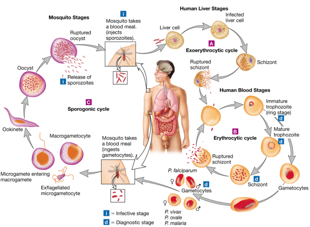
Vector competence is the intrinsic ability of anophe- line species or populations to allow the development of Plasmodium parasites from ookinete to infective sporo- zoites. When a mosquito takes an infectious blood meal from human, the gametocytes ingested begin their development in the midgut. The male gametocyte transform into eight microgametes after three rounds of mitosis, meanwhile the female gametocytes matures into macrogametes [4]. These cells fuse to form zygotes that thereafter change into ookinetes in the lumen of the intestine. The ookinetes then strive through the epithelium of the midgut and once in its basal side, transform into oocysts. The oocysts undergo several rounds of asexual multiplication (sporogony) leading to the production of thousands of haploid sporozoites in each oocyst. Mature occysts rupture and release sporo- zoites in the hemocoel, which immediately migrate to the salivary glands. The extrinsic incubation period of the parasite is about 14 days with the transition from ookinetes to mature oocysts having the highest dura- tion (about 10 days) [18, 19].

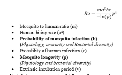
In mosquito host, Plasmodium face several immune- related bottlenecks deployed to prevent the success- ful transition from its early stage in the midgut to the sporozoite stage in the salivary glands [18]. The out- come of the parasite infection is reported to depend mainly on the mosquito-Plasmodium genetics adapta- tion [19, 20]. Another very important factor that influ- ences the above outcome is the compatibility of the duration of parasite development with the longevity of the mosquitoes [21, 22]. Only species in which Plas- modium reaches infective form are referred to as com- petent vectors and could ensure malaria transmission. The impact of vector competence on the transmission of malaria can be estimated using Ro (Fig. 1), the basic reproductive number developed by McDonald in 1957. The McDonald model gives the threshold for a disease to persist or spread (Ro greater than 1) or to disappear (Ro less than 1) [23]. The Ro represents the number of individuals in a susceptible human population that are expected to get infected via a mosquito bite when a sin- gle infected individual is present in the population [24, 25]. In the Ro equation, two parameters are related to vector competence: probability of mosquito infection (b)andmosquitolongevity(p)(Fig.1).Modificationsof the values of components of this equation for a given vector population will cause either an augmentation or reduction in the transmission dynamics of the disease, leading probably to a change in the epidemiological profile of the locality concerned. It was established that an increase in b will increase the Ro, whereas a decrease in p will cause the opposite [26].

Fig. 1 Basic reporductive number (Ro), Ross‐MacDonald model. In bold, parameters of the vectorial competence influenced by insecticide resistance

Insecticide resistance and malaria vector infectivity
to Plasmodium parasite


The rapid spread of insecticide resistance among malaria vectors accross endemic countries in the past dec- ade have raised several questions among which that of knowing what is its impact on mosquito permissiveness to Plasmodium? Only a limited number of studies have tried to elucidate this question [27–35]. These stud- ies compared P. falciparum infection rates in resistant Anopheline vectors with susceptible ones, either caught in the field or experimentally infected (Table 1).

Anopheles gambiae strain bearing kdr resistance allele (VgscL1014S) was found naturally more infected by sporozoites than the susceptible counterpart [27]. Similar findings were experimentally observed in the same spe- cies, as well as in Anopheles coluzzii [30, 32]. Contrary to kdr resistance, An. gambiae with ace-1 resistance allele did not differ from individuals that have the wild type allele (not conferring insecticide resistance) on infec- tion rate despite significantly higher oocyst prevalences were observed in the resistant strain [32]. More studies using field populations are needed to ascertain whether a lower longevity suspected by the author and/or other factors are involved.

Regarding metabolic resistance, recent breakthroughs in designing simple PCR-based assays to detect glu- tathione S-transferase (GST)-based and cytochrome P450-mediated resistance in An. funestus sensu stricto provided a unique opportunity to assess its impact on the mosquito’s ability to develop the parasites. The L119F- GSTe2 resistant genotypes of this species showed, in an experimental infection study, higher permissiveness to oocyst infections than susceptible ones [31]. Similarly, in naturally infected populations of the same species, homozygote L119F-GSTe2 genotypes were found more infected by sporozoites though no significant difference was found at the level of oocyst prevalence [28]. In other hands, Lo and Coetzee [36], infecting experimentally two selected sub-colonies of FUMOZ displaying different degree of pyrethroid resistance by Plasmodium berghei, found that the insecticide resistant colonies were less permissive to infection than the susceptible ones. No investigation has so far explored the relationship between P450s genes implicated in insecticide resistance and P. falciparum infection in An. funestus. Moreover, because of the absence of markers of metabolic resistance in An. gambiae sensu lato such studies are still lacking in these species.

Mosquito speciesType of experiment performedType of infectionInfection outcomeReferences
PrevalenceIntensity

An. gambiaea
kdr‐resistant strain compared to susceptible strainNaturalHigher sporozoite infection prevalence in resistant strains[27]
An. funestus s.saGSTe2‐resistant genotypes compared to susceptible genotypesNatural
No difference in oocysts infec‐ tion prevalence ns
Higher sporozoite infection prevalence in resistant strains

[28]

An. gambiae s.l.a
ace-1‐resistant strain com‐ pared to susceptible strain
Natural

Higher oocyst infection preva‐ lence in resistant strains

[29]
An. gambiae s.l.a
kdr‐resistant strain compared to susceptible strain
Natural
No difference in oocysts infec‐ tion prevalence ns

[29]

An. gambiaea
kdr‐resistant strain compared to susceptible strainExperimental Higher oocyst infection preva‐ lence in resistant strains Higher sporozoite infection prevalence in resistant strains
Higher oocyst load in the
resistant strains
Higher sporozoite infection load in the resistant strains
[30]
An. coluzziiakdr‐resistant strain compared to susceptible strainExperimental
Higher oocyst prevalence in the resistant strains
Higher sporozoite infection prevalence in resistant strains

Higher oocysts load in the resistant strains
Higher sporozoite load preva‐
lence in the resistant strains
[30]

An. funestus s.sa
GSTe2‐resistant genotypes compared to susceptible genotypes
Experimental

Lower oocyst infection preva‐ lence in homozygous resistant genotypess
Higher oocysts load inhomozygous and heterozy‐
gous resistant genotypess
[31]

An. gambiaeb

ace-1‐resistant strain com‐ pared to susceptible strain

Experimental

Higher oocyst prevalence infection in resistant strains No difference in sporozoite infection prevalencens

No difference in oocyst and sporozoite infection loadns
[32]

An. gambiaeb

kdr‐resistant strain compared to susceptible strain

Experimental

Higher oocyst infection preva‐ lence in resistant strains Higher sporozoite infection prevalence in resistant strains

Lower oocyst and sporozoite infection load in resistant
strains
[32]

An. gambiaeb

ace-1‐resistant strain com‐ pared to susceptible strain
Expérimental
Higher oocyst infection preva‐ lence in resistant strains

[33]

An. gambiaeb

kdr‐resistant strain compared to susceptible strain


Higher oocyst infection preva‐ lence in resistant strains

[33]
An. gambiaebace-1‐resistant strain com‐ pared to ace-1‐resistant strain exposed to insecticidesExperimental
Lower oocyst infection preva‐ lence in resistant strains

Lower oocyst infection load in resistant strains
[34]

An. gambiaea

kdr‐resistant strain compared to kdr‐resistant strain exposed to insecticide‐treated nets

Experimental

Lower oocyst infection preva‐ lence in resistant strains

Lower oocyst infection load in resistant strains
[35]
Table 1 Summary of studies evaluating the impact of insecticide resistance on malaria vectors susceptibility to P. falciparum
a: field strain; b: laboratory strain; ns: Non Significant; s: Significant; α: insecticide exposure to confirm resistance status probably post infection; β: insecticide exposure to confirm resistance status prior to infection

Impact of insecticide resistance on mosquito sialome

Bloodsucking arthropods, like mosquitoes, have evolved saliva containing a mixture of pharmacologically active molecules that help them counteract the hemostatis and inflammatory responses of the vertebrate host during bites, thus facilitating blood meal intake [37]. However, the activity of these molecules goes beyond the scope of ensuring blood meal success, as they possibly influ- ence the completion of Plasmodium development in the salivary gland of malaria vectors. Proteins secreted by

the salivary gland belong to several families (D7, mucin, gSG1, gSG2, gSG6 peptide, gSG7, cE5, 8.2-kDa, 6.2-kDa, etc.) [38] whose function include (1) cytoskeletal and structural activities (2) digestion, (3) circadian rythm and chemosensory, (3) immunity, (4) metabolism and other [39]. The development of insecticide resistance in malaria vectors is accompanied by physiological changes [26] that may affect the sialome composition with consequences on the vector competence. Few studies have investigated changes in the sialome in the insecticide resistant vectors [40, 41].

The secretory protein 100 kDa, which is encoded by Saglin (a cytoskeletal and structural gene present in An. gambiae salivary gland) was considered as the binding target of P. falciparum and P. berghei on salivary gland prior to penetration into the latter [42]. This protein was found down-regulated in ace-1 bearing An. gam- biae strain, suggesting an impact on the vector infectiv- ity to Plasmodium [43]. However, a recent study showed that the 100 kDa Protein is unevenly distributed on the salivary glands lobes. Its absence on the primary site of sporozoites occupancy in the salivary glands, the distal lateral lobes, implies that this protein may instead have a secondary role in the infection of the organ [44–46].

The D7 salivary family has been identified in malaria vectors among the most expressed proteins involved in the antihemostatic activity and probably in digestion of blood meal [47–50]. Elanga et al. [40] showed that two short forms of the D7 family genes (D7r3 and D7r4) are over-expressed in pyrethroid resistant An. funestus (L119FGSTe2), whereas almost all D7 genes are under- expressed in pyrethroid resistant An. gambiae (kdr, L1014F). A comparable observation was made in insec- ticide resistant Culex quinquefasciatus (ace-1 resistance) [51] as well as in two strains of Aedes aegypti (homozy- gotes resistant C1534 and G1016 kdr) [52]. These find- ings show that insecticide resistance mechanism may affect the sialome composition differently.

Several immune proteins such as the anti-microbial peptides cecropin and defensin were found in the saliva of mosquitoes [39, 53]. These immune proteins under- score the role of the salivary gland in the refractoriness of the Anopheles to infections [39, 53]. The small num- ber of studies that evaluated the impact of insecticide resistance alleles on salivary gland gene expression in mosquito vectors have not reported significant changes related to immune genes as compared with the suscepti- ble counterparts [41, 43, 51, 52], alluding that the resist- ant status to insecticide does not influence noticeably the immune component of the sialome. If these factors are indeed unchanged regardless of the mosquito allelic composition, nothing is known whether under infection the expression profile of these immune proteins will vary or not according to the mosquito genotype. Das et al. [39] and Djegbe et al. [51] demonstrated that salivary gland genes expression is influenced by blood meal intake and varies towards the period coinciding with the matura- tion of Plasmodium parasites in mosquitoes [54]. This evidence was not previously studied and should be taken into account in subsequent research works that aims at identifying differentially expressed genes of the salivary

gland and elucidating their impact on the malaria vector competence.

Impact of insecticide resistance on vector immunity

When the infectious blood meal reaches the midgut of the female Anopheles, the immune system is deployed to prevent infections [20]. In the midgut, P. falciparum faces the peritrophic membrane, a physical barrier developed to prevent infections. It also protects against the damag- ing effects of the human blood factors like antibodies and regulates several digestive enzymes [55, 56]. Enzymes such as trypsin 1 and 2, chymotrypsin, carboxypeptidase, aminopeptidase and serine protease are upregulated during digestion to cleave the large content of proteins in the blood meal [57–60]. These proteases are appar- ently involved in the elimination of Plasmodium infec- tions [61]. Three studies attempted to elucidate the effect of insecticide resistance on vectors’ immunity [62–64]. Mitri et al. [62], in a study evaluating genes implicated in the infectivity of An. coluzzii, demonstrated that the kdr– bearing para gene which carries mutations of the voltage- gate sodium channel (confering insecticide resistance) is not associated with infection but rather the ClipC9 gene directing the synthesis of Serine protease. This suggest that the effect of the resistant character on refractoriness to infection may be due to genes other than that involved in resistance to insecticides, and which happen to be linked to it. The Serine protease plays an important role in the activation of the three major immune signaling pathways in mosquitoes: Toll, Imd and JAK/STAT [20], which cause the release of antimicrobial peptides (AMPs) notably defensins, cecropins, attacin, gambicin and AgSTAT-A, effective against malaria parasites infections. Vontas et al. [63], using pyrethroid and organochlorine resistant An. gambiae strains, showed that defensin and cecropin are upregulated after pre-exposure to perme- thrin. This study sugggests that insecticide resistant mos- quitoes may be better equipped than susceptible ones to combat infections, but these two immune effectors alone may not be decisive in rendering the vector completely refractory to malaria infections as many other pathways activated concomitantly during parasitic invasion are altogether implicated in the outcome of a contamination [20].

In Culex pipiens which is vector of many pathogens including arboviruses [65], filarial worms [66], and protozoa [67], immune response was stimulated in an insecticide resistant field strain by injection of Lipopoly- sacharide (LPS) immune elicitor. As result, no difference was found in the expression of defensin and cecropin as compared to the control group; but only an increase in gambicin was recorded [68]. One point can be drawn from these results to infer what might happen in malaria vectors: Plasmodium infections may trigger the overex- presion of some immune factors while the other may have their expression either down regulated or unchanged.

The reactive oxygen species (ROS) produced by cellu- lar metabolism are another class of effectors of the innate immunity that can negatively affect malaria parasites [69, 70]. They kill the parasites through both lytic and melanization pathways [20]. Ingaham et al. [64] showed that An. coluzzii VK7 colony displaying kdr resistance mechanism, CYP6M6 and CYP6P3 metabolisers, had oxidoreductase overexpressed after sub-lethal exposure to deltamethrin, suggesting that this species could be more refractory to Plasmodium infection. At this point, it is necessary to verify whether under natural conditions, insecticide-resistant Anopheles mosquitoes will display an overexpression of ROS or not.

Cellular immune responses are carried out by varous type of hemocytes that eliminate pathogens by phago- cytosis, lysis and melanization [20]. Organochlorines and organophosphate were found to affect differently the hemocytes abundance including granulocytes in the insect Rhynocoris kumarii [71]. In mosquitoes, studies are needed to ascertain the impact of insecticide resist- ance on cellular immunity and the resulting effect on the infectivity of resistant vector to malaria parasite. Regard- ing melanization of pathogens, it is lead by the phe- noloxidase (PO) produced by Oenocytoids [72, 73] and is regulated by serine protease inhibitors. In field-caught C. pipiens resistant to insecticide through an increase in detoxification (esterase) and target site mutation (ace- 1), PO expression was equal to that of susceptible group [74], suggesting that some genes associated with immu- nity might not be affected by insecticide resistance char- acter in mosquitoes. No studies have verified the effect of insecticide resistance on PO in malaria vectors.

Impact of insecticide resistance on commensal intestinal microbiota of malaria vectors
Bacteria, fungi and viruses colonize the gut, salivary glands and reproductive organs of the mosquitoes. These microorganisms are mainly acquired from the environne- ment and its composition is largely influenced by its aquatic breeding sites [75, 76]. In addition, the microbi- ota composition is highly dynamic, varying greatly with localities and seasons [77–79]. These variations of micro- biota composition within field mosquitoes may partly explain the variability in infection levels in the field [80].

Mosquito microbiota has great potential for impeding the transmission of malaria by altering vectorial capac- ity [81]. Also, the microbiota is capable of influencing the biology of the host such as altering its immunity, nutri- tion, digestion, vectorial competence, reproduction, and insecticide resistance [82–87]. With the growing concerns about the rapid spread of insecticide resistance in Anopheles mosquitoes, some studies have explored the functions of the mosquito’s gut microbial communities in the development of resistance. For example, distinct microbita populations were found associated with organ- ochlorine resistance in An. arabiensis [86] and Anoph- eles albimanus [88]. Similarly, an association between specific microbiota and intense pyrethroid resistance was reported in An. gambiae [89] and Anopheles ste- phensi [90], suggesting a microbiota-mediated insecticide resistance mechanism. Dieme et al. [91] suggested that changes in the feeding behaviour of insecticide resistant vectors may lead to higher microbial diversity. This diver- sity could modify the repertoire of protective bacteria against pathogen infections and/or that of their enhanc- ers, with consequences on the vectorial competence [9, 92]. Recently, Bassene et al. [93] showed that, in the spe- cies An. gambiae and An. funestus, the microbiota was signifanctly different between P. falciparum-infected and non-infected samples, although the resistance status of these mosquitoes was not evaluated. More refined stud- ies are needed to characterize the microbial communities harboured by the insecticide resistant malaria vectors. Also the contribution of microbiota against other fac- tors to the vectorial competence of insecticide resistant malaria vectors remains to be investigated.

Impact of insecticide resistance on the longevity of malaria vectors
Mosquito longevity is a determinant factor for parasite maturation and could influences malaria transmission [94, 95]. In fact, the extrinsic incubation of Plasmodium in its hematophageous host is about 11–14 days. There- fore, only mosquitoes whose lifespan is long enough could allow the complete development of the malaria parasite to the sporozoite infective stage and participate in the transmission of the disease. With the emergence and spread of insecticide resistance [7], many investiga- tions were undertaken to gain knowledge of the effect of this phenomenon on the vectors’ longevity and so on its potential epidemiological impact.

So far, studies on the impact of insecticide resistance on malaria vectors longevity have focused on four spe- cies: An. gambiae, An. arabiensis, An. coluzzii and An. funestus. Globally, the findings revealed a pleitropic effect of insecticide resistance on mosquito lifespan [33, 96–108] (Table 2). The majority of studies (10/14) which used laboratory strains showed that pyrethroid resistant An. funestus and An. gambiae live longer than suscepti- ble ones [100, 106]. Of the studies including field strains, a longer life span was reported in organochlorine and pyrethroid resistant An. funestus strains [104, 105]. In contrast, a shorter life span was observed in An. gambiae strain resistant to organochlorine. It was reported that pre-exposure to insecticide in a manner micmicing field exposure to insecticides, affects the longevity of insecti- cide resistant An. gambiae strain [97], and that delayed mortality observed in the vectors may be dependent on resistance intensity [98]. This later observation indi- cates that findings obtained with laboratory colonies are to be taken with caution given that they may not reflect exactly what is oberved on the field [26]. Nevertheless, such studies remain important as they contribute to the understanding of the potential mechanisms affecting the vectors’ longevity [109, 110], notably resource-based trade-off and oxidative stress.

Study speciesOrigin of strainsClass of insecticidesResistance mechanism(s)
Pre-Exposurea/Exposureb to insecticide

Effect on longevity

References

An. gambiae s.l

Field

PY

Not available

Deltamethrin and permethrina

RR longevity > SS longevity

[97]

Field
PY & OCkdrNot appliedRR longevity < SS longevity[96]
FieldPY
Not available
Permethrin (Net) bRRe longevity=RRne
[98]
LaboratoryPY & OCkdr & P450
Permethrin (Hut‐net) b
LaboratoryPYNot availableDeltamethrinb
RR longevity < SS longevity[99]
LaboratoryPYkdr, P450 and esteraseNot applied
RR longevity > SS longevity[100]
LaboratoryOCkdrDDTa
RR longevity = SS longevity[33]
CAace‐1
Bendiocarba
RR longevity > SS longevity
LaboratoryOCkdr, GSTe, P450 & EsNot appliedRR longevity = SS longevity[101]
LaboratoryOCGSTe, P450 & EsNot appliedRRb longevity > RRs[102]
LaboratoryOCGSTe, P450, Es;kdrNot appliedRR longevity < SS longevity[103]
DDTa
RRe longevity = RRne
PermethrinaRRe longevity < RRne
DeltamethrinaRRe longevity < RRne
Malathion
RRe longevity < RRne
An. funestusField
OC
GSTe2
Permethrin (Net) bRR longevity > SS longevity[104]
FieldPY & OCGSTe2Not appliedRR longevity > SS longevity[105]
LaboratoryPYNot available Not applied RR longevity > SS longevity[106]
LaboratoryPY & CAP450-a
Not applied RR longevity = SS longevity[107]
LaboratoryPY & CAP450-aNot applied RR longevity = SS longevity[108]
P450-bRR longevity = SS longevity
P450-a/P450-bRR longevity = SS longevity
Table 2 Summary of studies assessing the impact of insecticide resistance on the longevity of malaria vectors Impact of insecticide resistance on malaria vector competence: a literature review
PY: Pyrethroid; OC: Organochlorine; CA: Carbamate; GSTe: Glutathion S-transferase; Es: Esterase; kdr: Knock down resistance; P450: Cytochrome P450 monoxygenase; RR: resistant strain, SS: susceptible strain; RRe: resistant strain exposed to insecticide; RRne: resistant strain non-exposed to insecticide; RRb: Resistant strain fed on blood; RRs: Resistant strain fed on sugar; > more; < less; = equal

Resource based trade-off is an evolutionary ecology concept that states that when environmental constraints lead to the augmentation of resources to one biological trait, other traits will have their energy budget reduced [111]. Accordingly, when mosquitoes adopt the detoxi- fication mechanism to prevent the effect of insecticide, an increased production of detoxifying enzymes follows and is maintained by the additional resources deployed for the function. Otali et al. [100] have demonstrated

that metabolism and longevity of insecticide resist- ant An. gambiae are lower than that of the susceptible strain. Moreover, they showed that the resistant strain has higher Reactive Oxygen Species (ROS), which are factors determining oxydative stress. In fact, the ROS are multifunctional molecules produced by cells of all organisms during normal metabolism [112, 113]. They have been pointed out as key aging factor in other organ- isms including Anopheles [114]. Therefore, mosquitoes that develop the capacity to cope with oxydative stress are likely to live longer. Oliver and Brooke [103] in an experiment evaluating the effect of oxidative stress on the longevity of both An. arabiensis and An. funestus bear- ing respectively kdr and Cytochrome P450 mechanisms demonstrated that these species live longer, and that Cytochrome P450 activity seems more protective against oxydative stress.

Rivero et al. [26] proposed the potential effect of dif- ferent detoxifying enzymes on vector longevity. For example, Glutathion S-Transferase is considered to pro- tect against oxydative stress. Confirming this point, a longer lifespan implicating Glutathion S-Tranferase in An. funestus was revealed with and without exposure to insecticide [104, 105]. In contrast, monoxygenase, known to be associated with an increase in oxydative stress has not led, as expected, to reduced longevity in An. funes- tus [108]. More studies using field populations and mic- micing field conditions are necessary to ascertain the full impact of insecticide resistance on longevity of the malaria vector.

Conclusion

The need for a comprehensive understanding of the impact of insecticide resistance on malaria vector com- petence is unquestionable. The current state of knowl- edge is not only insufficient but also contradictory to draw a definitive conclusion. A tendency nevertheless emerges from findings that insecticide resistance may increases the infectivity of malaria vectors to Plasmo- dium, thus their vector competence. This is possibly due to changes in the expression of some genes notably those involved in blood-feeding and the immunity. Addition- ally, microbiota communities vary in the resistant mos- quitoes as compared to the susceptible counterparts. The actual effect of these changes in the course of infection and their impact on the infectivity of malaria vectors to P. falciparum is still to be investigated. Finally, the longevity of the vectors is not always affected by insecticide resist- ance mechanisms. It is worth noting that, studies using vectors displaying metabolic resistance were under- represented because molecular markers to diagnose this character were developped only recently, especially in An. funestus. Malaria vectors that bear metabolic resistant mechanism are, on an ecological immunology point of view, expected to have a number of biological functions impaired, including immunity. If established, this situa- tion may cause them to become less refractory to Plas- modium infection. Taking advantage of recent advances in the genomics, transcriptomics and molecular charac- terization of insecticide resistance, more refined studies can now be undertaken to fill knowledge gaps regarding the effect of insectide resistance on key determinants of vectorial competence and subsequently predict changes in the epidemiology of malaria in a context of insecticide resistance escalation.

DDT Dichloro‐diphenyl‐trichloroethane GST Glutathione S‐transferases
kdr Knock down resistance
LPS Lipopolysacharide

PO Phenoloxidase
Vgsc Voltage gate sodium channel

Author contributions

PFS, EEN, MT, MMS, CN wrote the manuscript. TD and PFS prepared the figures and tables. CN and CW acquiered the funding. All the authors revised the manuscript. All authors read and approved the final manuscript.

Funding

The authors acknowledge the financial support from the Wellcome Trust, UK, through the International Intermediate Fellowship (220720/Z/20/Z) granted to Cyrille NDO.

Availability of data and materials

Not applicable.

Declarations
Ethics approval and consent to participate

Not applicable.

Consent for publication

Not applicable.

Competing interests

The authors declare no competing interests.

References

Accepted: 4 January 2023

1. WHO. A global brief on vector‐borne diseases. Geneva, World Health Organization; 2014.

2. Gelband H, Panosian CB, Arrow KJ. Saving lives, buying time: economics of malaria drugs in an age of resistance. 2004.

3. WHO. World malaria report 2021. Geneva, World Health Organization; 2021.

4. Phillips M, Burrows J, Manyando C. Malaria. Nat Rev Dis Primers. 2017;3:17050.

5. Cox FE. History of the discovery of the malaria parasites and their vec‐ tors. Parasit Vectors. 2010;3:5.

6. Sinka ME, Bangs MJ, Manguin S, Rubio‐Palis Y, Chareonviriyaphap T, Coetzee M, et al. A global map of dominant malaria vectors. Parasit Vec‐ tors. 2012;5:69.

7. Riveron JM, Tchouakui M, Mugenzi L, Menze BD, Chiang M‐C, Wondji CS. Insecticide resistance in malaria vectors: an update at a global scale. In Towards malaria elimination‐a leap forward. IntechOpen; 2018.

8. Elliott R, Ramakrishna V. Insecticide resistance in Anopheles gambiae Giles. Nature. 1956;177:532–3.

9. Sheldon BC, Verhulst S. Ecological immunology: costly parasite defences and trade‐offs in evolutionary ecology. Trends Ecol Evol. 1996;11:317–21.

10. Barnes KG, Weedall GD, Ndula M, Irving H, Mzihalowa T, Hemingway
J, et al. Genomic footprints of selective sweeps from metabolic resist‐ ance to pyrethroids in African malaria vectors are driven by scale up of insecticide‐based vector control. PLoS Genet. 2017;13: e1006539.

11. Donnelly MJ, Corbel V, Weetman D, Wilding CS, Williamson MS, Black WC. Does kdr genotype predict insecticide‐resistance phenotype in mosquitoes? Trends Parasitol. 2009;25:213–9.

12. Weill M, Lutfalla G, Mogensen K, Chandre F, Berthomieu A, Berticat C,
et al. Insecticide resistance in mosquito vectors. Nature. 2003;423:136–7.

13. Djogbénou L, Labbé P, Chandre F, Pasteur N, Weill M. Ace‐1 duplica‐ tion in Anopheles gambiae: a challenge for malaria control. Malar J. 2009;8:70.

14. Hemingway J, Hawkes NJ, McCarroll L, Ranson H. The molecular basis of insecticide resistance in mosquitoes. Insect Biochem Mol Biol. 2004;34:653–65.

15. Riveron JM, Irving H, Ndula M, Barnes KG, Ibrahim SS, Paine MJ, et al. Directionally selected cytochrome P450 alleles are driving the spread activity of midgut aminopeptidases. Arch Insect Biochem Physiol.

1990;15:149–63.

  1. Dana AN, Hong YS, Kern MK, Hillenmeyer ME, Harker BW, Lobo NF, et al. Gene expression patterns associated with blood‐feeding in the malaria mosquito Anopheles gambiae. BMC Genomics. 2005;6:5.
  2. Billingsley PF, Hecker H. Blood digestion in the mosquito, Anopheles stephensi Liston (Diptera: Culicidae): activity and distribution of trypsin, aminopeptidase, and alpha‐glucosidase in the midgut. J Med Entomol. 1991;28:865–71.
  3. Ribeiro JM. A catalogue of Anopheles gambiae transcripts significantly more or less expressed following a blood meal. Insect Biochem Mol Biol. 2003;33:865–82.
  4. Vijay S, Rawal R, Kadian K, Singh J, Adak T, Sharma A. Proteome‐wide analysis of Anopheles culicifacies mosquito midgut: new insights into the mechanism of refractoriness. BMC Genomics. 2018;19:337.
  5. Mitri C, Markianos K, Guelbeogo WM, Bischoff E, Gneme A, Eiglmeier K, et al. The kdr‐bearing haplotype and susceptibility to Plasmodium falciparum in Anopheles gambiae: genetic correlation and functional testing. Malar J. 2015;14:391.
  6. Vontas J, Blass C, Koutsos AC, David JP, Kafatos FC, Louis C, et al. Gene expression in insecticide resistant and susceptible Anopheles gambiae strains constitutively or after insecticide exposure. Insect Mol Biol. 2005;14:509–21.
  7. Ingham VA, Brown F, Ranson H. Transcriptomic analysis reveals pronounced changes in gene expression due to sub‐lethal pyrethroid exposure and ageing in insecticide resistance Anopheles coluzzii. BMC Genomics. 2021;22:337.
  8. Hamer GL, Kitron UD, Brawn JD, Loss SR, Ruiz MO, Goldberg TL, et al. Culex pipiens (Diptera: Culicidae): a bridge vector of West Nile virus to humans. J Med Entomol. 2008;45:125–8.
  9. Morchón R, Bargues MD, Latorre JM, Melero‐Alcíbar R, Pou‐Barreto C, Mas‐Coma S, et al. Haplotype H1 of Culex pipiens implicated as natural vector of Dirofilaria immitis in an endemic area of Western Spain. Vector Borne Zoonotic Dis. 2007;7:653–8.
  10. Kimura M, Darbro JM, Harrington LC. Avian malaria parasites share congeneric mosquito vectors. J Parasitol. 2010;96:144–51.
  11. Vézilier J, Nicot A, Lorgeril J, Gandon S, Rivero A. The impact of insecti‐ cide resistance on Culex pipiens immunity. Evol Appl. 2013;6:497–509.
  12. Molina‐Cruz A, DeJong RJ, Charles B, Gupta L, Kumar S, Jaramillo‐ Gutierrez G, et al. Reactive oxygen species modulate Anopheles gambiae immunity against bacteria and Plasmodium. J Biol Chem. 2008;283:3217–23.
  13. Kumar S, Christophides GK, Cantera R, Charles B, Han YS, Meister
    S, et al. The role of reactive oxygen species on Plasmodium mel‐ anotic encapsulation in Anopheles gambiae. Proc Natl Acad Sci USA. 2003;100:14139–44.
  14. James RR, Xu J. Mechanisms by which pesticides affect insect immu‐ nity. J Invertebr Pathol. 2012;109:175–82.
  15. Hillyer JF, Strand MR. Mosquito hemocyte‐mediated immune responses. Curr Opin Insect Sci. 2014;3:14–21.
  16. Lavine MD, Strand MR. Insect hemocytes and their role in immunity. Insect Biochem Mol Biol. 2002;32:1295–309.
  17. Cornet S, Gandon S, Rivero A. Patterns of phenoloxidase activity in insecticide resistant and susceptible mosquitoes differ between labora‐ tory‐selected and wild‐caught individuals. Parasit Vectors. 2013;6:315.
  18. Wang Y, Gilbreath TM 3rd, Kukutla P, Yan G, Xu J. Dynamic gut microbi‐ ome across life history of the malaria mosquito Anopheles gambiae in Kenya. PLoS ONE. 2011;6: e24767.
  19. Gimonneau G, Tchioffo MT, Abate L, Boissière A, Awono‐Ambéné PH, Nsango SE, et al. Composition of Anopheles coluzzii and Anopheles gambiae microbiota from larval to adult stages. Infect Genet Evol. 2014;28:715–24.
  20. Akorli J, Gendrin M, Pels NA, Yeboah‐Manu D, Christophides GK, Wilson MD. Seasonality and locality affect the diversity of Anopheles gambiae and Anopheles coluzzii midgut microbiota from Ghana. PLoS ONE. 2016;11: e0157529.
  21. Krajacich BJ, Huestis DL, Dao A, Yaro AS, Diallo M, Krishna A, Xu J, Lehmann T. Investigation of the seasonal microbiome of Anopheles coluzzii mosquitoes in Mali. PLoS ONE. 2018;13: e0194899.
  22. Sandeu MM, Maffo CGT, Dada N, Njiokou F, Hughes GL, Wondji CS. Seasonal variation of microbiota composition in Anopheles gambiae

and Anopheles coluzzii in two different eco‐geographical localities in

Cameroon. Med Vet Entomol. 2022;36:269–82.
80. Rosenberg R. Malaria: some considerations regarding parasite produc‐

tivity. Trends Parasitol. 2008;24:487–91.
81. Cansado‐Utrilla C, Zhao SY, McCall PJ, Coon KL, Hughes GL. The micro‐

biome and mosquito vectorial capacity: rich potential for discovery and

translation. Microbiome. 2021;9:111.
82. Rodgers FH, Gendrin M, Wyer CAS, Christophides GK. Microbiota‐

induced peritrophic matrix regulates midgut homeostasis and prevents systemic infection of malaria vector mosquitoes. PLoS Pathog. 2017;13: e1006391.

83. Song X, Wang M, Dong L, Zhu H, Wang J. PGRP‐LD mediates A. stephensi vector competency by regulating homeostasis of microbiota‐induced peritrophic matrix synthesis. PLoS Pathog. 2018;14:e1006899.

84. Romoli O, Gendrin M. The tripartite interactions between the mosquito, its microbiota and Plasmodium. Parasit Vectors. 2018;11:200.

85. Barletta ABF, Trisnadi N, Ramirez JL, Barillas‐Mury C. Mosquito midgut prostaglandin release establishes systemic immune priming. iScience. 2019;19:54–62.

86. Barnard K, Jeanrenaud A, Brooke BD, Oliver SV. The contribution of gut bacteria to insecticide resistance and the life histories of the major malaria vector Anopheles arabiensis (Diptera: Culicidae). Sci Rep. 2019;9:9117.

87. Martinson VG, Strand MR. Diet‐microbiota interactions alter mosquito development. Front Microbiol. 2021;12: 650743.

88. Dada N, Sheth M, Liebman K, Pinto J, Lenhart A. Whole metagenome sequencing reveals links between mosquito microbiota and insecticide resistance in malaria vectors. Sci Rep. 2018;8:2084.

89. Omoke D, Kipsum M, Otieno S, Esalimba E, Sheth M, Lenhart A, et al. Western Kenyan Anopheles gambiae showing intense permethrin resist‐ ance harbour distinct microbiota. Malar J. 2021;20:77.

90. Soltani A, Vatandoost H, Oshaghi MA, Enayati AA, Chavshin AR. The role of midgut symbiotic bacteria in resistance of Anopheles stephensi (Dip‐ tera: Culicidae) to organophosphate insecticides. Pathog Glob Health. 2017;111:289–96.

91. Dieme C, Rotureau B, Mitri C. Microbial pre‐exposure and vectorial competence of Anopheles mosquitoes. Front Cell Infect Microbiol. 2017;7:508.

92. Gabrieli P, Caccia S, Varotto‐Boccazzi I, Arnoldi I, Barbieri G, Comanda‐ tore F, et al. Mosquito trilogy: microbiota, immunity and pathogens, and their implications for the control of disease transmission. Front Microbiol. 2021;12: 630438.

93. Bassene H, Niang EHA, Fenollar F, Dipankar B, Doucouré S, Ali E, et al. 6S Metagenomic comparison of Plasmodium falciparum‐infected and noninfected Anopheles gambiae and Anopheles funestus microbiota from Senegal. Am J Trop Med Hyg. 2018;99:1489–98.

94. Garrett‐Jones C, Shidrawi GR. Malaria vectorial capacity of a population of Anopheles gambiae: an exercise in epidemiological entomology. Bull World Health Organ. 1969;40:531–45.

95. Ferguson HM, Maire N, Takken W, Lyimo IN, Briët O, Lindsay SW, Smith TA. Selection of mosquito life‐histories: a hidden weapon against malaria? Malar J. 2012;11:106.

96. Nkahe DL, Kopya E, Djiappi‐Tchamen B, Toussile W, Sonhafouo‐Chiana N, Kekeunou S, et al. Fitness cost of insecticide resistance on the life‐traits of a Anopheles coluzzii population from the city of Yaoundé, Cameroon. Wellcome Open Res. 2020;5:171.

97. Msangi G, Olotu MI, Mahande AM, Philbert A, Kweka EJ. The impact of insecticide pre‐exposure on longevity, feeding succes‐ sion, and egg batch size of wild Anopheles gambiae s.l. J Trop Med. 2020;2020:8017187.

98. Hughes A, Lissenden N, Viana M, Toé KH, Ranson H. Anopheles gambiae populations from Burkina Faso show minimal delayed mortality after exposure to insecticide‐treated nets. Parasit Vectors. 2020;13:17.

99. Viana M, Hughes A, Matthiopoulos J, Ranson H, Ferguson HM. Delayed mortality effects cut the malaria transmission potential of insecticide‐ resistant mosquitoes. Proc Natl Acad Sci USA. 2016;113:8975–80.

100. Otali D, Novak RJ, Wan W, Bu S, Moellering DR, De Luca M. Increased production of mitochondrial reactive oxygen species and reduced adult life span in an insecticide‐resistant strain of Anopheles gambiae. Bull Entomol Res. 2014;104:323–33.

  1. Oliver SV, Brooke BD. The effect of elevated temperatures on the life his‐ tory and insecticide resistance phenotype of the major malaria vector Anopheles arabiensis (Diptera: Culicidae). Malar J. 2017;16:73.
  2. Oliver SV, Brooke BD. The effect of multiple blood‐feeding on the lon‐ gevity and insecticide resistant phenotype in the major malaria vector Anopheles arabiensis (Diptera: Culicidae). Parasit Vectors. 2014;7:390.
  3. Oliver SV, Brooke BD. The role of oxidative stress in the longevity and insecticide resistance phenotype of the major malaria vectors Anoph- eles arabiensis and Anopheles funestus. PLoS ONE. 2016;11: e0151049.
  4. Tchakounte A, Tchouakui M, Mu‐Chun C, Tchapga W, Kopia E, Soh PT,
    et al. Exposure to the insecticide‐treated bednet PermaNet 2.0 reduces the longevity of the wild African malaria vector Anopheles funestus but GSTe2‐resistant mosquitoes live longer. PLoS ONE. 2019;14:e0213949.
  5. Tchouakui M, Riveron JM, Djonabaye D, Tchapga W, Irving H, Soh Takam P, et al. Fitness Costs of the glutathione S‐transferase epsilon 2 (L119F‐ GSTe2) mediated metabolic resistance to insecticides in the major African malaria vector Anopheles funestus. Genes (Basel). 2018;9:645.
  6. Okoye PN, Brooke BD, Hunt RH, Coetzee M. Relative developmental and reproductive fitness associated with pyrethroid resistance in the major southern African malaria vector, Anopheles funestus. Bull Entomol Res. 2007;97:599–605.
  7. Tchouakui M, Riveron Miranda J, Mugenzi LMJ, Djonabaye D, Wondji MJ, Tchoupo M, et al. Cytochrome P450 metabolic resistance (CYP6P9a) to pyrethroids imposes a fitness cost in the major African malaria vector Anopheles funestus. Heredity (Edinb). 2020;124:621–32.
  8. Tchouakui M, Mugenzi LMJ, Wondji MJ, Tchoupo M, Njiokou F, Wondji CS. Combined over‐expression of two cytochrome P450 genes exac‐ erbates the fitness cost of pyrethroid resistance in the major African malaria vector Anopheles funestus. Pestic Biochem Physiol. 2021;173: 104772.
  9. Remick D. Measuring the costs of reproduction. Trends Ecol Evol. 1992;7:42–5.
  10. Kirkwood TB, Rose MR. Evolution of senescence: late survival sacrificed for reproduction. Philos Trans R Soc Lond B Biol Sci. 1991;332:15–24.
  11. Garland T Jr. Trade‐offs. Curr Biol. 2014;24:R60–1.
  12. Rada B, Leto TL. Oxidative innate immune defenses by Nox/Duox family NADPH oxidases. Contrib Microbiol. 2008;15:164–87.
  13. Sumimoto H. Structure, regulation and evolution of Nox‐family NADPH oxidases that produce reactive oxygen species. FEBS J. 2008;275:3249–77.
  14. Monaghan P, Metcalfe NB, Torres R. Oxidative stress as a mediator of life history trade‐offs: mechanisms, measurements and interpretation. Ecol Lett. 2009;12:75–92.

by Dr Rostand Idriss Tchana, Pharmacist

Resume

Drug delivery system have been developed past years improved and tried to improve the best security and care for the patients. There is a lot of technologies involved in this process as the industry raised several investments in nanomaterials (Shuang et al., 2017).

The impact of these different improvements in patient care leads to specific intelligent biomaterials designed to release in response to either physical, chemical or biological triggers. The specific use of this DDS leads to spatiotemporal control and deeply optimize the drug effects (Carlos M. et al., 2019).

Mostly the strategies are using drug conjugated to the precarrier (prodrug), in an inactive state who’s degraded in-vivo and deliver on target the active agents.  The unique structure of the produgs showed self-assembly properties with nanoparticles with nanoparticles through chemical modifications. Also several advantages been present on them including high efficiency, amelioration of drug availability, drug release controllability and less tendency to agglomerate after encapsulation (  Luo C. et al., 2014). After consideration, we can devide the DDS responsive to some stimuli into two categories addressed as internal (endogenous) responsive DDS and external (exogeneous) responsive DDS.

 It’s really obvious for us as pharmacists to have a bibliographical study on the topic and check the late advances in the topic and what improvements can be done to have an optimistic innovation and best care for the patients.

Introduction

Drug delivery systems and galenic formulation technologies are the most important concerns of the pharmaceutical industry. It’s really important to have the drug delivered to the targeted site and avoiding non-targeted sites relying on adverse effects and also toxicity.

The latest advancements in DDS technologies Currently, lead to improvements in nanomedicines in cancer and several pathologies such as lymphoma, leukemia, inflammation etc. Different technologies merged in this field as liposomes and micelles (Liu et al., 2019), built up as polymer nanoparticles, with the significant advancement of nanotechnology, a variety of nanomedicines have been developed as polymer nanoparticles (Shuang et al., 2017), inorganic nanocrystals (Yang G. et al., 2017), biomimetic nanomaterials (Ouyang J. et al, 2018).

The different elements integrated into the Stimuli-responsive DDS can be divided into two big types depending on the stimuli type used : internal stimuli using the tumor micro-environment (TMO) (e.g., Redox environment, pH, enzymes and hypoxic etc.)  and external stimuli not in the tumor environment but induced by the therapist voluntarily (e.g., light, thermo, ultrasound, and magnetism field, etc.) (Anel Xie et al., 2020).

The prodrug (drugs conjugated to the precarrier), is inactive after administration and can be degraded in vivo to gain the required efficiency, this with a simple modification of its structure. This system gives several advantages as higher bioavailability, better efficiency, control on the release (target), and less risk of agglomeration after encapsulation ( Luo C. et al, 2014).

It is now obvious and relevant to discuss the importance of prodrug-based nanomedicines specified in  the sensitive respond to certain stimuli for drug delivery systems (DDS).

Context

As we’re in an evolving world where the purpose is to give the best care to the patients with an optimistic balance risks/benefits, drug delivery on the target site with minimal toxicity to the organs that are not supposed to be targeted remains a significant challenge in modern drug delivery technologies. Past years, advancements such as combinatory chemistry, biotechnology, gene-based drugs, and high throughput screening provide several new molecular entities for various diseases, but with many of these drugs, the optimal treatment is only possible if sufficient delivery of the compounds to the target site of action. Also, treatments with biological drugs, such as DNA and proteins, may suffer from deficient drug delivery to the target cells (V.P. Torchilin, 2018).

Nanomedicine drug delivery has recently received tremendous attention in the treatment of various diseases such as breast  and ovarian cancer (Sankha and al. 2022). Drug delivery to the target tissues such as head and neck cancer and even infected regions of the body, brain and back eye segment is challenging due to the high difficulty to reach the right concentration amount of the active principles in those regions (Ishwor Poudel and al. 2022). Therefore, the need for localized and targeted delivery is becoming increasingly important in reducing toxicity and side effects, increasing the  drug efficacy with a decrease of dose and even medicines costs. The nonspecific delivery of drugs and tissue biodistribution and rapid drug metabolism or excretion from the body makes the drug action less efficient and requires increased dose witch causes a real challenge for drug design and the pharmaceutical industry.

Thus the understanding of molecular aspects associated with different pathologies has led to deep understanding of endogenous and exogeneous material that can be exploited to devise “stimuli responsive” material that can be targeting specific sites of application into the body. These are new targets of the pharmaceutical industry regarding chronic diseases  that can be exploited need to develop new DDS and mainly SRD are the main target in new pharmaceutical strategies and the industry is investing more and more in these concepts.

Stimuli responsive drugs

Stimuli responsive drugs are one of the latest innovation in therapeutic and drug delivery and may constitute an ideal and innovative approach to have more results with less side effects in several treatments.

Stimuli responsive drugs are drugs made out of material that can modify reversibly or not their physical properties in response to an internal or external stimuli, causing change in their particles properties such as dimensions, structure and interactions, which leads to rearrangements on their aggregation state where the responsiveness is supplied from a part of the material.

There are different therapeutical applications for SRD, most used are in oncology where the latest innovation were the use of MSR-DDSs are a fine touch and offer a good platform for co-delivering of agents and constitute a really interesting spot to avoid multidrug resistance (Jia R and al., 2021).

There is different classification to SDR due to the type of material they’re made of and also the type of stimuli they’re responsive to : Internal or external stimuli responsive drugs depending on the location of the stimuli element chosen, and depending of the type of stimuli we can have pH, light, ultrasound or redox stimuli used. Therefore we can manage to match them into different ways to represent SDR (fig. 1) where we do have in blue external stimuli responsive drugs and in red internal stimuli responsive drugs.

Figure 1 Schematic representation of exogenous and endogenous stimuli-responsive vesicular systems. Source: From (N.d.).

Some Definitions

Drug delivery system :

Drug delivery system are the system used to deliver drugs / active agents to the target sites of the pharmacological agents.

To reach their target in the body, drugs can be inserted following different types of route depending on the active principle and the target routes of the drug itself. These routes can be classified as depending on their “starting point” ( Laura Elizabeth Lansdowne, Technology Networks / Drug Discovery, December 2020).

Thus we do have the following delivery routes that can be used to our drugs to reach targets in the body (fig 2) :

Buccal drug delivery

Buccal drug delivery corresponds to the administration of a drug through the buccal mucosa (lining of the cheek) (Jinsong Hao et al. , 2003) . This delivery route avoids gastrointestinal degradation process and first-pass effect (rapid drug uptake and metabolism into inactive compounds by the liver), there is a real “challenging” barrier to drug absorption through this route, especially for larger biopharmaceuticals – delivery is currently limited to small molecule drugs with lipophilic properties as they can readily cross the membrane (Flavia Laffleur, Drug Development and Industrial Pharmacy, 2014 ).  The use of buccal route for drug delivery is often for extended-release drug delivery (Drug release on an extended time period with a controlled process), A deep connection and adhesion/attachment to the mouth mucosa are mostly preferred. Different formulations have been developed for buccal delivery including; tablets, gels, lozenges and patches (Jinsong Hao et al. , 2003).

Nasal drug delivery

Nasal drug delivery involves the delivery of a drug through the nose with a connection with the nasal cavity. Nasal spray medications are usually used for the treatment ofpathologies affecting the upper respiratory tract (e.g., nasal congestion, allergic rhinitis) ( Karim Amighi, Galenic Pharmacy course, 2021 – 2022). We can also in some circumstances use this this route for a systemic target for example regarding the delivery of small molecules drugs – e.g., the migraine medication olmatriptan (Zomig®) (CBIP, consultation le 26/07/2022).The great vascularization of this mucosa helps to increase access to blood circulation for drugs and avoidance of first-pass metabolism.

Ocular drug delivery 

Ocular drug delivery is a tricky route for the scientists ant the patients as well. This is due to the unique anatomy and physiology of this organ having a static, dynamic and metabolic ocular barriers impede the absorption of drugs through the eye. This organ can use different targets of drugs delivery with a specific approach. Different approaches and innovative works helped passing  challenges of delivering drugs to ocular tissues through identification of different transporters (efflux and influx transporters) and modification of drugs technology formulation to target these transporters  in the eye and modifying drugs to target these transporters (Gaudana et al. 2010).

Oral drug delivery

The most common route used for drug is the oral drug delivery, several key points justifies this use as a way of drug administration as non-invasive nature, ease of use, cost-effectiveness and the highly absorptive properties of the gastrointestinal (GI) tract.  are Oral drug delivery is by far the most well-known and often the preferred route of drug administration due to its non-invasive nature, ease-of-use, cost-effectiveness, and the highly absorptive properties of the gastrointestinal (GI) tract ( CBIP, 2022). Several conditions are required for the oral delivery to be successful, the aqueous solubility of the drug compound in the GI system as well to make sure if there is any modification to the drug technology to be added for increase of the bioavailability. This route requires particular attention of caregivers and health practitioners for specific patients populations  including; pediatric, geriatric and those with cognitive impairment.

Pulmonary drug delivery

For this delivery route, the inhalation through the mouth and into the airways is required. These can be used for treating local diseases in the lungs. The effects of this drug technology use can be for a local or systemic effect.  Last advancements in science helped to move forward with pulmonary drug delivery as a potential route of administration for systemic diseases regarding the vast absorption potential due to the large absorptive surface area and the great permeable membrane of the alveolar region. This route is also a great space due to the  unaffected value  by dietary and interpatient variability ( Labiris NR et al., 2003).

Sublingual drug delivery

This route of drug delivery is refereeing for drug administration under the tongue, then absorption into the bloodstream via the tongue’s ventral surface and the mouth floor is observed. of the mouth ( Narang et al. , 2010) . The fast absorption of sublingual route helps to set rapid drug effect. Sublingual drug delivery system also avoids hepatic first‐pass metabolism effect. Somehow, patient’s compliance can be affected because of interaction with talking, eating and drinking where they can cause disruption. In addition, absorption and consequently efficacy of the drug is reduced by smoking due to vasoconstriction of the vessels, therefore use in smokers is not recommended (Hao Zhang et al., 2002).

Transdermal drug delivery

Transdermal drug delivery refers to a DDS method for systemic effect by applying the formulation directly onto intact skin ( of delivering a drug systemically by applying a formulation onto intact skin ( Alkilani et al., 2015). The trajected  way used by the drug with this route is first penetration through the stratum corneum and then progression through deep epidermis and dermis and absorption via microcirculation. The non-invasive characteristic of this route can help for patients unconscious or suffering from regurgitations ( Praunitz et al., 2004).

Vaginal/anal drug delivery

Anal and vaginal drug have a faster onset of action compared to the oral route and a higher bioavailability. Their use could be for local or systemic effect depending on the targeted pathology. First-pass metabolism is avoided and no interactions with gastrointestinal disturbances is observed (Wolfson et al., 2000).

Figure 2 Different drug delivery routes Lilley et al., Pharmacology and the nursing process 2011

Stimuli Responsive drugs :

Stimuli responsive drugs are drugs that responds to a specific trigger to deliver their cargo on the specific site targeted. Their use is mostly to overcome adverse drug effect and optimize efficiency depending on the route of administration ( Rahim MA et al., 2021). We can summarize the different effect of the SRD strategies on the figure below.

Figure 3 Stimuli responsive drugs, Xiao Fu et al. 2018

Internal Stimuli responsive drugs

Internal stimuli responsive drugs are drugs made out of smart biomaterials that respond to triggers present in their microenvironment  such as pH or specific enzymes ( Wells CM et al., 2019).

External Stimuli responsive drugs

External stimuli responsive drugs concerns drugs that are made with smart biomaterials responding to triggers present out of their microenvironment such as electromagnetic field, light or even acoustic stimulaton ( Wells CM et al., 2019).

Multi-Stimuli responsive drugs

Multi-Stimuli responsive drugs are drugs made with biomaterials that responds to double or triple triggers depending on the formulation. These materials are engineered to have high sensibility to the surrounding physiological environments and release the encapsulated cargo with a deep specificity to their target (Xiao Fu et al, 2018).

Therapeutical Applications

In DDS, stimuli responsive drugs are used for different pathologies but mostly tumors are the one targeted,the use of these materials can be shortened in the table below regarding the use of nanocarriersin vesicular DDS ( table 1)

Table 1 Different Vesicular systems under clinical trial with applied filters : active ( not recruiting), Interventionnal Studies ( Clinical trials) at clinicaltrials.gov Other sources : H. He, L. Liu, E. E. Morin, M. Liu, A. Schwendeman. Survey of clinical translation of cancer – nanomedicines – lessons learned from successes and failures Acc Chem Res 2019 52(9):24452461 AC Anselmo, S Mitragotri Nanoparticles in the clinic : any update Bioeng Transl Med 2019 ,4 ( 3) : e10143 DJ Crommelin, P Van Hoogevest G Storm The role of liposomes in clinical nanomedicine development What Now ? now What? J Controlled Rel, 2020 318:256263

Vesicular systemCommercial products or clinical trialsApplications (approved)
DoxilDoxil/FDA approved (1995)Breast cancer, Ovarian cancer
Doxorubicin liposomes (Pegylated)Caelyx/EMA approved (1996)HIV-associated Kaposi’s sarcoma
Daunorubicin liposomes (Pegylated)DaunoXome/FDA approved (1996)HIV-associated Kaposi’s sarcoma
Paclitaxel liposomesLipusu/Approved in China (2003)Metastatic gastric cancer
Cytarabine liposomesDepocyt/Approved in USA (2006)Leukemia
Paclitaxel micellesGenexol-PM/Approved in Korea (2007)Breast cancer
Mifamurtid liposomesMEPACT/EMA approved (2009)Osteosarcoma (Primary) following surgery
Irinotecan liposomes (Pegylated)Onivyde/FDA approved (2015)Metastatic pancreatic cancer (secondary)
Cytarabine:Daunorubicin, (5:1) liposomesVYXEOS CPX-351/FDA approved (2017)Acute myeloid leukemia
Propofol liposomesDiprivan/FDA approved (1989)Induction of anesthesia
Amphotericin-B liposomesAmBisome/FDA approved (1997)Cryptococcal meningitis in HIV-infected patients
Systemic fungal infections
Visceral leishmaniasis in immunocompromised patients
Verteporfin liposomesVisudyne/FDA approved (2000)Age-related macular degeneration
Doxorubicin liposomes (Pegylated)Doxil/Caelyx: 241#Solid malignancies, ovarian, breast cancer, luekemia, lymphomas, prostate, metastatic or liver cancer
Daunorubicin liposomesDaunoxome: 48#Leukemia
Doxorubicin liposomesMyocet: 22#Breast cancer, lymphoma and ovarian cancer
Paclitaxel liposomesLipusu: 1#Breast and nonsmall cell lung cancer
Cytarabine liposomesDepocyt: 11#Solid tumors, Lymphoma
Mifamurtide liposomesMepact: 1#Osteosarcomas
Vincristine liposomesMarqibo: 149#Cancers including lymphoma, brain, leukemia, melanoma
Irinotecan liposomes (Pegylated)Onivyde: 7#Solid Malignancies, breast cancer, pancreatic cancer, sarcomas
Cytarabine:Daunorubicin (5:1) liposomesVyxeos: 2#Leukemias
Propofol liposomesDiprivan: 19#General anesthesia in morbidly obese patients, open heart surgery, spinal surgery
Amphotericin-B liposomesAmbisome: 7#Invasive fungal infections
Verteporfin liposomesVisudyne: 1#Macular degeneration
Cisplatin (Pegylated) liposomes (Lipoplatin)Phase-3Nonsmall cell lung cancer
Paclitaxel liposomes (EndoTAG-1)Phase-3Breast cancer

Pancreatic adenocarcinoma
Doxorubicin liposomes HER2 targeted (MM-302)Phase-3Breast cancer
Doxorubicin liposomes (thermally sensitive) (ThermoDox)Phase-3Hepatocellular carcinoma
VCL-1005 plasmid liposomes (Allovectin-7)Phase-3Melanoma
MUC1 antigen liposomes
(Tecemotide)
Phase-3Nonsmall cell lung cancer
Paclitaxel micelles (NK105)Phase-3Breast cancer
Cisplatin micelles (NC-6004)Phase-3Pancreatic cancer
Irinotecan and floxuridine liposomes (CPX-1)Phase-2Colorectal cancer
Bis-neodecanoate diaminocyclohexane platinum liposomes (Aroplatin)Phase-2Colorectal cancer
Lurtotecan liposomes (OSI-211)Phase-2Ovarian cancer
SN38 liposomes (LE-SN38)Phase-2Colorectal cancer
Paclitaxel liposomes (LEP-ETU)Phase-2Breast cancer
Annamycin liposomesPhase-2Acute lymphocytic leukemia
Tretinoin liposomes (Atragen)Phase-2Kidney cancer
Doxorubicin liposomes (glutathione Pegylated) (2B3101)Phase-2Solid tumors and brain cancer
Protein Kinase N3 siRNA liposomes  (Atu027)Phase-2Pancreatic cancer
p53 plasmid liposomes (SGT-53)Phase-2Pancreatic cancer, glioblastoma
SN38 micelles (NK012)Phase-2Breast cancer
Doxorubicin micelles (SP1049C)Phase-2Advanced esophagus or gastroesophageal junction adenocarcinoma
Docetaxel micelles (CPC634)Phase-2Ovarian cancer

Categories

Internal Stimuli Responsive drugs :

As presented in the definition part, internal stimuli responsive drugs refers to drugs made with biomaterials or nanocarriers having the ability to respond specific triggers located in their micro-environment. These triggers can be physiological ( enzyme, redox, thermic …) or linked to the specific pathology targeted  ( Xiao Fu et al. 2018).

Enzyme-responsive drug delivery system :

Enzyme-responsive DDS refers to system that has modification of physicochemical properties after enzymatic actions, this may lead to the liberation of the active agents and obtention of the therapeutic effect wanted (Sabya Sachi et al., 2020). Many enzymes like lipase, protease, trypsin, glycosidase, phospholipase, oxidoreductase etc. are selected in this therapeutic strategy and for several pathologies such as cancer therapies ( Nguyen et al. 2015). The process is simple and can be summarized on the picture below

Figure 4 Enzyme-responsive nanomaterials for drug delivery and diagnostics; a) polymer-based nanoparticles can be covalently modified with drugs through an enzyme-cleavable linker so that the enzyme activity triggers drug delivery in the tissue of interest; b) polymer-stabilized liposomes can be loaded with drugs, whose degradation can be programmed to be triggered by an enzyme. c) Inorganic particles can be used for diagnostic when the activity of the target hydrolase controls the assembly or disassembly of the nanoparticles, which turn in changes the nanoparticle solution

Here are several uses of enzymes-responsive technologies in DDS :

  • MPP-Responsive, polymeric systems

High matrix metalloproteinases (MMPSs) concentrations have been identified and associated with many cancerous cells. This special type of enzyme could selectively cleave peptide linkages in between nonterminal amino acid sequences ( Van Rijt S. H. et al. 2015). MMP2-sensitive siRNA delivering system development, with self-assembling and  copolymeric technology of the polyethylene glycol-peptide-polyethylenimine-12, along with dioleoyl-snglycero-3-phosphoethanolamine (PEG-pp-PEI-PE), is referred as one of the latest innovation. The linker was based on an octapeptide, GPLGIAGQ, which is high sensitive to MMP2 with big response. From this process high tumor targeting of the developed system merged (Zhu L. et al. 2014). A different study showed the use of N-(3-aminopropyl) methacrylamide (APM) and acrylaminde (AAM) to synthesize copolymeric nanocapsules with MMP-responsive peptide cross-linkers to cargo BSF and VEGF (Wen J. et al, 2014). The design of novel MMP2 sensitive nanoparticles were used in a recent work, these designed from copolymeric TPGS3350-pp-PLA along with TPGS-folate to deliver anticancer drugs (Pan J. et al, 2018). An MMP 8-responsive, polymeric hydrogel was developed from a diacrylate-containing polyethylene glycol-based moiety, along with a cysteine-terminated peptide cross-linker (CGPQG↓IWGQC). The encapsulation  system used for the hydrogels was made with , and antibacterial peptide KSL and they were used to target chronic periodontitis and peri-implantitis (Guo J. et al, 2019).

  • Esterase-responsive, polymeric systems

This system relies on the ester bond cleavage to the targeted drug Several  esterase-responsive, polymeric systems have been developed lately by researchers, we will present some here. The development of the PAMAM-based, polymeric dendrimer is focused to his link to PTX via succinate bond. This PTX/PAMAM G4 dendrimers were readily hydrolyzed by esterase and then release the free drug, this system helps an optimization of internalization in effective cell and toxicity reduction. Upon conjugation with PEG, these systems revealed an enhanced action which could be explored to develop more anticancer drugs ( Khandare J.J. et al., 2006).  For pathologies such as inflammation, a novel twin-drug system, i.e., Dexamethasone-Diclofenac, merged with a system including esterification reaction and then encapsulated into polylactide (PLA) nanoparticles. In vivo studies of the were done using esterase systems. Results shows that esterase hydrolysis enhanced the drug release rate, and the synergistic activity of the two anti-inflammatory drugs led to higher inhibition of the TNF-α level than the free drug ( Assali M. et al., 2018).

  • Cathepsin B.

The use of the  tetrapeptide linkage Gly-Phe-Leu-Gly is one of the common viewed in the enzyme-responsive design, polymeric systems, this is justified by the easy cleavage by cathepsin B, an lysosomal enzyme with  expression in tumor tissues (GIANASI E. ET AL, 2002). The development of a PEGylated gemcitabine-containing with cathepsin B responsive action was made up to target tumor microenvironments (Zhang C. , 2017). Another study with  a peptide macromonomer was designed from BIM and cathepsin B substrate, which was then incorporated into copolymeric deblock system of DEAEMA/BMA and DEAEMA. This shown a great and promising result in the intracellular delivery of peptides (Kern H. B. et al, 2017).

Swelling controlled drug delivery system :

“Swelling-controlled drug delivery system” refers to therapeutical devices where the release is controlled by the swelling step. This can also be used for devices where the swelling step has a high importance or plays an important role on the mass transport process. (e.g., drug dissolution, drug diffusion, and polymer dissolution) (Siepmann J. et al., 2012).

Most of swelling-controlled DDS are based on hydrophilic polymers like the hydroxypropyl methylcellulose ( Colombo P. et al, 2008; Escudero JJ et al, 2010). We can see on the figure 5 the process on the polymer swelling. From dry state (non-swollen state) where the polymer has a high density and the mobility of the macromolecules is restricted to the swollen state where due to hydratation we can see a relaxation on the polymer chains and increase of volume and mobility of the macromolecules. Thus the physical changes on the polymer drives the control of the release rate of the drug.

Figure 5 Schematic presentation of a drug-loaded polymeric network in the non-swollen state (top) and upon liquid penetration into the system (bottom). If the polymer is soluble in water, an erosion front separates the bulk fluid from the swollen polymeric network. Crosses represent dissolved drug molecules, black diamonds non-dissolved particles. ( Juergen Siepmann et al., 2011)

pH-responsive drug delivery system :

pH  is a major factor regarding the DDS for any drug administrated into the body, the delivery of a drug must manage that the physiological needs at the targeted sites  matches deeply  to the different organs targeted and the tissues or cellular compartment  properties. Thus pH is a great parameter on Stimuli responsive drugs and other DDS. The dependency of the therapeutic efficacity to the control in time and location of delivery are one of the points raised by this DDS (Zhu YJ. Et al, 2015).

Under normal physiological states, pH varies on different sites and remains more or less stale depending on the location site and the organs (table 2).

Table 2 pH values in physiological conditions (Yongxu Mu et al. 2021)

SitepH value
Plasma7,38 – 7,42
Saliva6,0 – 7,0
Gastric Juice1,0 – 3,5
Bile7,8
Pancreatic juice8,0 – 8,3
Small Intestinal fluid5,5 – 7,0
Lysosome4,5 – 5,0
Golgi Apparatus6,0 – 6,7

Different strategies are used on the pH responsive DDS, depending on the material used and the targeted sites; literature divided them into four big responses modes ( Yongxu Mu et al., 2021) :

  • physical dissociation
  • chemical bond cleavage -resulting nanoparticle dissociation
  • chemical bond cleavage promoting payload release
  • nanoparticle swelling

The process can easily be observed on the figure bellow

Figure 6 Release mode of acid-sensitive pH-responsive polymeric micelles (a) Physical dissociation; (b) Chemical bond cleavage-resulting nanoparticle dissociation; (c) Chemical bond cleavage-promoting payload release; (d) Nanoparticle swelling ( C. Huang et al, 2010; M. Kanamala et al., 2016)

Redox-Responsive DDS :

The use of Redox-responsive DDS is mostly for polymer technologies. In this process the disulfide is cleaved by a high concentration of GSH, an abundant thiol present in cell cytoplasm. Its function is mostly reduction in biochemical processes ( Thambi et al., 2016). GSH concentrations varies deeply depending on the tissue where they’re present (normal or pathological) and the pathological state of the patient. In some pathologies like neurodegenerative diseases, liver diseases, stokes, seizures or diabetes we can have an increase rate of GSH (Estrella et al., 2006). Thus this difference of redox in the environment is also present in tumor cells and is definitely useful to build up DDS based on redox-responsive nanomaterials (Schafer et al., 2001).

Here is a figure representing the use of nanocarriers in the redox-responsive mechanism ( (figure7).

Figure 7 Schematic illustration of nanocarriers in the redox-responsive mechanism. The drug-carrying nanocarriers enter the cell through the endocytosis process. Glutathione (GSH) reduces the disulfide bonds after approaching the cytosol and subsequently erupts and releases the drug. ( Fleige et al. 2012)

Thermo-Responsive drugs :

Thermo-responsive system are mostly used in DDS for therapeutical strategies; they can be used for both diagnostic and treatment (Son H. et al; 2020). Mostly dehydration is the main process leading to drug release on the thermos-sensitive process in DDS.  The NCs response to the external temperature change modify the polymer chain and this can then lead to drug release (Liu et al., 2016). Mostly tumor cells do have a higher temperature ( 40 – 42°C ) than normal cells (37°C). Thus the release of the payload is obtained by the contact of the specified tissues with higher temperature ( Liu et al., 2016). 

Two different approaches can be used for DDS with thermos-responsive NCs  :

  • NCs responsive to high temperature drug release :

We can have for example in this part the polycaprolactone ( N-Isopropyl acrylamide) (PNIPAM)  which displays tunable properties and a reduction of the LCST with an increase of the PAA content. Its use can be applied to various switching of DDS where he responses to small pH changes. Their use showed a cell viability of 20% at 40°C versus a viability of 40% at 37°C for treatments using this technology ( Son H. et al.; 2020).

The second approach involving external stimuli of higher temperature will be developed on the following point.

Glucose-triggered drug delivery system :

The use of hydrogels in DDS and other therapeutic applications as soft biomaterials have increased lately in different applications. The deep resemblance to hydrogels and  the extracellular matrix (ECM) conditions  plus their ability to load the drugs and other bioactive molecules made hydrogels promising candidates for the development of smart DDS (Allan S. Hoffman, 2012). Hydrogels that respond to specific stimuli have attracted the attention of researchers because of their potential for space, temporal and ondemand delivery options. Their use can be sorted in different ranges of application from sensing, biotech to tissue engineering ( Heidi R. Culver et al., 2017). The research groups of Chang-Li and Xiaoyu Liu showed dual-sensitive nanogels that can be used in improvement of  oxidation-responsive and glucose triggered hydrogels for insulin delivery (Li C et al., 2019).

The same type of preparation using the oxidation-responsive hydrogels used a four-arm polyethylene glycol (PEG) containing reactive oxygen cleavable acrylic bonds supported by phenylboronic ester pendant groups was used in M. Zang work (Figure 8.a). These hydrogels were formed mostly with hydrogels hydrophilic PEG and a fraction of hydrophobic linker molecules. Their mechanical strength was an asset to a macroscopic gel structure under culture conditions, and less toxicity appeared with LO2 cells in vitro. Cleavage of hydrogels and the release of payloads is achieved only in the presence of reactive oxygen stimuli such as hydrogen peroxide (H2O2). Glucose oxidase (GOx), is an enzyme causing glucose’s oxidation to gluconolac-tone and produces H2O2 is encapsulated into the hydrogels. (Shivaprasad M. et al., 2018). GOx incorporation has bestowed the hydrogels with a glucose responsive nature. The use of hydrogels with GOx and FITC-insulin is tremendous in the demonstration og glucose triggered release of insulin  under in vitro conditions. The release of encapsulated insulin (FITC-insulin) in the presence of external H2O2 and glucose was monitored using fluorescence measurements and the loss of gel integrity was measured by rheology and visual disintegration of the hydrogels (Figure 8.b). The release of encapsulated insulin was found to be dependent on the concentration of triggering molecules used (either H2O2 or glucose) and a faster release of insulin along with degradation of hydrogels was observed at high concentrations of stimuli molecules. Strikingly, the GOx encapsulated hydrogels were found to be responsive to the pathological glu- cose concentration (20 μM) and were triggered to release the insulin under in vitro conditions(Shivaprasad M. et al., 2018).

GOx encapsulated oxidation sensitive PEG hydrogels have showed several properties as high selectivity, sensitivity and cytocompatibility this ended up with hydrogels giving temporal on-demand release of insulin with monitoring of insulin release depending on the needs. These strategies can be used in several diabetics cases as foot ulcers. The ability of hydrogels to release payloads in the presence of H2O2 stimuli further extends the suitability in the treatment of ROS associated diseases and tissue engineering.

Figure 8 Glucose triggered insulin releasing hydrogels. (a) Schematic representation of synthesis, encapsulation of GOx, FITC-insulin into hydrogels and glucose/H2O2 triggered release through degradation. (b) Photo- graphs showcasing the time-dependent release of  FTIC-insulin and degradation in the presence of H2O2 and Glucose. Zhang M., C. Song, F. Du and Z. Li, ACS Appl. Mater. Interfaces, 2017 , 9, 25905 Copyright 2017 American Chemical Society.

External Stimuli Responsive drugs :

Thermo-responsive Nanomedicines drugs :

The second approach used in DDS with thermo-responsive NCs is the use of carriers that burst drug release in response to external triggers increasing the temperature in the drug carrier. This alters the thermo-sensitive DDS and end-up to a drug release on the targeted site (Khoee S. et al., 2018).  This process may include for example PPy in amphiphilic di-block copolymer for tumor treatment. PPy has a photothermal property which leads to the promotion of drug release from the micelles (Son H. et al; 2020).

Magnetic-responsive Nanomedicines drugs

Lately the use of magnetic-responsive materials in the microelectronics, biomedical applications, microfluidics and chemistry fields increased deeply. Magnetism is an external non-invasive method of activation that has attractive capabilities because it can be controlled in a temporal and spatial  (Mahdi Karimi et al., 2015). The use of DDS with Magnetic stimuli response are considered as best option for the design of efficient drug delivery and lesser interaction than some other stimuli responsive DDS,e.g. pH, ultrasound and light (Mophatra A. et al., 2019).

Photo-responsive DDS :

Several elements as non-invasiveness, inexpensiveness, and practicability leaded to the use of light interest for DDS as an exogenous stimulus. Its use for prodrug-based nanomedicines as an external stimuli gave many advantages over other internal stimuli following the ease of handling, precise control of the time and location of treatment ( Mura S. et al, 2013). The use of  light-responsive smart prodrug-based nanosystems using ultraviolet (UV), visible light, and near-infrared (NIR) ( figure 9.)  have been intensively applied for non-invasive and controlled on-demand drug release against cancer (Schroeder A. et al, 2012 ; Yang G. et al,2016 ; Jia S. et al., 2018).  

Due to the consequences on structural transitioning modifications with UV light (300-380nm) on polymers with special structures ( as  O-nitrobenzyl, pyrene, spiropyran, and azobenzene), it is widely used in prodrug release ( Jia S. et al, 2018 ; Azagarsamy M.A. et al, 2012 ; Tong R. et al, 2012).  We do have for example Liu’s group work using a photocaged linkage to form prodrugs-based shell cross-linked (SCL) which were composed of P(CL-g-CPT)-b-P(OEGMA-co-MAEBA)-CPT and PCL-b-P(OEGMA-co-MAEBA-co-FA) amphiphilic diblock copolymers. This using a photocaged CPT prodrug enhancing cleavage and drug release on the micelles under UV irradiations ( Angel Xie et al., 2020).

Figure 9  General presentation of light responsive polymers applied to NIR ( B. Sana et al., 2022)

Some Materials used in DDS :

The huge demand for adapted and innovative processes in DDS raised more interests than ever in SRD and implementation of latest improvement in drug engineering and  nanotechnologies moved to the next generation of drugs with less side effects and huge efficiency.

In fact different materials are used in SRD formulation, depending on their payload and the specific receptors targeted; It’s important for this process to keep stability through the delivery process and maximum reduction of exogeneous interactions ( food, poly-medication, other medical tools etc.).

We will focus on the type of stimuli to categorize the different SRD, thus we have :

  • pH responsive DDS 

Depending on the type of disease, there is a wide range of materials pH-sensitive that can be used in DDS.  The engineering of  hydrogels for their physicochemical properties as stiffness, degradation and porosity helps to directly act with influence on the fates of the encapsulated cells including their migration, proliferation, differentiation, and communication. Furthermore, stimuli-responsive hydrogels could improve the in vivo therapeutic efficacy and also provide new therapeutic pathways ( Wang L. et al., 2018).

Thermos-responsive materials

  • Light-responsive materials
  • Redox-responsive materials and electroactive polymers
  • Magnetic-responsive nanomaterials

Challenges in DDS :

In conclusion, the nano-tech aided engineering novel polymeric carriers are hugely growing in various types of drug delivery applications. With key scientific advances, massive research efforts are being made with a particular focus to construct DDS that can respond to the physiochemical-based surrounding changes that are either external or internal in a sophisticated manner with high-level control and precision. In this paper, we summarized the uniqueness of stimuli-responsive polymeric carriers and their potentialities for targeted drug delivery applications. Considering suitable examples, an array of stimuli-responsive DDS, for instance, redox-responsive, pH-responsive, temperature-responsive, photo/light-responsive, magnetic-responsive, ultrasound-responsive, and electrical-responsive DDS, and/or all-in-all dual/dual/multi-responsive DDS (combination or two or more from any of the above) have been discussed. For easy understanding and to highlight the work behavior of different DDS, schematic illustrations are also included by giving the focus on chemistry aspects, release behavior, and drug delivery applications. Aside from the significant advancement in the biomedical/pharmaceutical at large and DDS arena, in particular, there are still many concerns that remain to be addressed. For instance, biocompatibility, biodegradability, non-toxicity, and safer elimination of the smart carriers from the biological system are some of the significant limiting factors that one needs to be considered prior to designing DDS. Furthermore, to induce fast responsiveness, the size of the carrier also plays a critical role. Nanosized carriers are being developed with comprisable mechanical characteristics, which is one of the important features required for biomedical-based applications. Besides processing drawbacks, administrative issues to get approval to use the designed polymeric-based smart DDS is another major bottleneck in the field. To avoid all these limitations, future investigations should be directed to introduce novel technologies to design DDS using polymeric materials with inherent properties such as biocompatibility, biodegradability, and non-toxicity. In conclusion, many polymer-based stimuli-responsive carriers offer substantial potential for biomedical applications. Thus, the external/internal stimuli-responsive carriers hold great promise for nanomedicine in the coming future.

Discussion :

Stimuli-responsive materials are a smart choice for the development of DDS meanwhile their use in pharmaceutical industry required adaptability and efficiency in the different targeted pathologies. The use of these materials the past years improved in several therapies as tumors, chronic inflammation, diabetes  and even diagnosis or anesthesia  procedures.

Access to a plethora of physical, chemical and biological stimuli have resulted in the design and development of smart drug delivery systems with controlled spatiotemporal drug delivery ( Aida Lopez et al., 2022).

 In our work we discussed a few selected examples of stimuli DDS based on pH, glucose, enzyme, light, magnetic field and ultrasound triggers under in vitro conditions, and an example of NIR-triggered in vivo used in photo responsive DDS.

Despite great progresses observed in smart drug delivery systems for various diseases using strategies with diverse stimuli triggers, there is still a need for next generation stimuli-responsive drug delivery systems and devices with more precise and on-demand delivery. Importantly, most of the reported stimuli-responsive drug delivery systems and devices are tested under either in vitro or in cellulo settings, but not under in vivo conditions involving real pathological situations and also interactions with several exogenous components as food or other drugs co-administrated to patients. In spite of the recent progress in stimuli-respon- sive smart delivery devices, there is a plenty of room for the development of next-generation multifunctional smart drug delivery systems and devices that are biocompatible and biodegradable, in addition to being spatiaotem- poral, target specific, for on-demand delivery.

Bibliography

  1. Alkilani, A.Z.; McCrudden, M.T.C.; Donnelly, R.F. Transdermal Drug Delivery: Innovative Pharmaceutical Developments Based on Disruption of the Barrier Properties of the Stratum Corneum. Pharmaceutics 2015, 7, 438-470.
  2. Allan S. Hoffman, Hydrogels for biomedical applications, Advanced Drug Delivery Reviews, Volume 64, Supplement, 2012, Pages 18-23, ISSN 0169-409X, https://doi.org/10.1016/j.addr.2012.09.010.
  3. B. Sana, A. Finne-Wistrand, D. Pappalardo, Recent development in near infrared light-responsive polymeric materials for smart drug-delivery systems, Materials Today Chemistry, Volume 25, 2022, 100963, ISSN 2468-5194,
  4. C. Huang, et al. Synthesis of polyhalo acridones as pH-sensitive fluorescence probes Bioorg. Med. Chem. Lett., 20 (15) (2010), pp. 4665-4669
  5. CBIP 2022 updates
  6. Colombo P, Santi P, Siepmann J, Colombo G, Sonvico F, Rossi A, Strusi OL (2008) Swellable and rigid matrices: controlled release matrices with cellulose ethers. In: Augsburger LL, Hoag SW (eds) Pharmaceutical dosage forms: tablets, vol 2: rational design and formulation, 3rd edn. Informa Healthcare, New York
  7. Cui D., Huang J., Zhen X. et al. A semiconducting polymer nano-prodrug for hypoxia-activated photodynamic cancer therapy. Angew Chem Int Ed. 2019; 58: 5920-5924
  8. Culver HR, Clegg JR, Peppas NA. Analyte-Responsive Hydrogels: Intelligent Materials for Biosensing and Drug Delivery. Acc Chem Res. 2017 Feb 21;50(2):170-178. doi: 10.1021/acs.accounts.6b00533. Epub 2017 Feb 7. Erratum in: Acc Chem Res. 2018 Oct 16;51(10):2600. PMID: 28170227; PMCID: PMC6130197.
  9. Dai Y., Xiao H., Liu J., et al. In Vivo multimodality imaging and cancer therapy by near-infrared light-triggered trans-platinum pro-drug-conjugated upconverison nanoparticles. J Am Chem Soc. 2013; 135: 18920
  10. Escudero JJ, Ferrero C, Jimenez-Castellanos MR (2010) Compaction properties, drug release kinetics and fronts movement studies from matrices combining mixtures of swellable and inert polymers. II. Effect of HPMC with different degrees of methoxy/hydroxypropyl substitution. Int J Pharm 387:56–64
  11. Fleige E, Quadir MA, Haag R. Stimuli-responsive polymeric nanocarriers for the controlled transport of active compounds: concepts and applications. Adv Drug Deliv Rev. 2012 Jun 15;64(9):866-84. doi: 10.1016/j.addr.2012.01.020. Epub 2012 Feb 11. PMID: 22349241.
  12. Freya Q. Schafer, Garry R. Buettner, Redox environment of the cell as viewed through the redox state of the glutathione disulfide/glutathione couple, Free Radical Biology and Medicine, Volume 30, Issue 11, 2001, Pages 1191-1212, ISSN 0891-5849, https://doi.org/10.1016/S0891-5849(01)00480-4.
  13. Gaudana, R., Ananthula, H.K., Parenky, A. et al. Ocular Drug Delivery. AAPS J 12, 348–360 (2010). https://doi.org/10.1208/s12248-010-9183-3
  14. Gianasi E., Buckley R.G., Latigo J., Wasil M., Duncan R. HPMA copolymers platinates containing dicarboxylato ligands. Preparation, characterisation and in vitro and in vivo evaluation. J. Drug Target. 2002;10:549–556. doi: 10.1080/1061186021000072456.
  15. Guan X., Chen Y., Wu X., Li P. and  Liu Y. Enzyme-responsive sulfatocyclodextrin/prodrug supramolecular assembly for controlled release of anti-cancer drug chlorambucil Chem Commun. 2019; 55: 953-956 https://doi.org/10.3390/pharmaceutics7040438
  16. Ishwor Poudel, Manjusha Annaji, Robert D. Arnold, Virendra Gajbhiye, Amit K. Tiwari, R. Jayachandra Babu, Chapter 3 – Vesicular nanocarrier based stimuli-responsive drug delivery systems, Editor(s): Virendra Gajbhiye, Kavita R. Gajbhiye, Seungpyo Hong, Stimuli-Responsive Nanocarriers, Academic Press, 2022, Pages 61-86, ISBN 9780128244562,
  17. Jia R, Teng L, Gao L, Su T, Fu L, Qiu Z, Bi Y. Advances in Multiple Stimuli-Responsive Drug-Delivery Systems for Cancer Therapy. Int J Nanomedicine. 2021 Feb 25;16:1525-1551. doi: 10.2147/IJN.S293427. PMID: 33658782; PMCID: PMC7920594.
  18. Jia S., Fong W.-.K., Graham B., Boyd B.J Photoswitchable molecules in long-wavelength light-responsive drug delivery: from molecular design to applications. Chem Mater. 2018; 30: 2873-2887
  19. Jinsong Hao & Paul W. S. Heng (2003) Buccal Delivery Systems, Drug Development and Industrial Pharmacy, 29:8, 821-832, DOI: 10.1081/DDC-120024178
  20. Karim Amighi, Galenic Pharmacy course PHARJ404, Academic Year 2020 – 2021
  21.  Kern H.B., Srinivasan S., Convertine A.J., Hockenbery D., Press O.W., Stayton P.S. Enzyme-Cleavable Polymeric Micelles for the Intracellular Delivery of Proapoptotic Peptides. Mol. Pharm. 2017;14:1450–1459. doi: 10.1021/acs.molpharmaceut.6b01178.
  22.  Khandare J.J., Jayant S., Singh A., Chandna P., Wang Y., Vorsa N., Minko T. Dendrimer versus linear conjugate: Influence of polymeric architecture on the delivery and anticancer effect of paclitaxel. Bioconjugate Chem. 2006;17:1464–1472. doi: 10.1021/bc060240p.
  23. Khoee, S.; Karimi, M.R. Dual-Drug loaded Janus graphene oxide-based thermoresponsive nanoparticles for targeted therapy. Polymer 2018, 142, 80–98
  24. Labiris NR, Dolovich MB. Pulmonary drug delivery. Part I: physiological factors affecting therapeutic effectiveness of aerosolized medications. Br J Clin Pharmacol. 2003 Dec;56(6):588-99. doi: 10.1046/j.1365-2125.2003.01892.x. PMID: 14616418; PMCID: PMC1884307.
  25. Laura Elizabeth Lansdowne, Technology Networks / Drug Discovery, December 2020
  26. Li C , Liu X , Liu Y , Huang F , Wu G , Liu Y , Zhang Z , Ding Y , Lv J , Ma R , An Y , Shi L . Glucose and H2O2 dual-sensitive nanogels for enhanced glucose-responsive insulin delivery. Nanoscale. 2019 May 9;11(18):9163-9175. doi: 10.1039/c9nr01554j. PMID: 31038150.
  27. Ling X., Tu J., Wang J. et al. Glutathione-Responsive Prodrug Nanoparticles for Effective Drug Delivery and Cancer Therapy. ACS Nano. 2019; 13: 357-370
  28. Liu D, Yang F, Xiong F, Gu N. The Smart Drug Delivery System and Its Clinical Potential. Theranostics. 2016 Jun 7;6(9):1306-23. doi: 10.7150/thno.14858. PMID: 27375781; PMCID: PMC4924501.
  29. Luo C, Sun J, Sun B, He Z. Prodrug-based nanoparticulate drug delivery strategies for can- cer therapy. Trends Pharmacol Sci 2014;35(11):556–66.
  30. M. Kanamala, W.R. Wilson, M. Yang, B.D. Palmer, Z. Wu Mechanisms and biomaterials in pH-responsive tumour targeted drug delivery: a review Biomaterials, 85 (2016), pp. 152-167
  31. Mahdi Karimi, Parham Sahandi Zangabad, Amir Ghasemi and Michael R Hamblin
    Smart External Stimulus-Responsive Nanocarriers for Drug and Gene Delivery Smart External Stimulus-Responsive Nanocarriers for Drug and Gene Delivery chapter 4 Magnetic-responsive nanocarriers; Published November 2015 • Copyright © 2015 Morgan & Claypool Publishers Pages 4-1 to 4-6
  32. McDaniel J.R., Bhattacharyya J., Vargo K.B. et al. Self-assembly of thermally responsive nanoparticles of a genetically encoded peptide polymer by drug conjugation. Angew Chem Int Ed. 2013; 52: 1683-1687
  33. Mohapatra A, Fujiwara T, Harris MA, LeVine DA, Ghimire M, Morshed BI, Jennings JA, Bumgardner J, Haggard WO, Mishra SR. Magnetic Stimulus Responsive DDS Based on Chitosan Microbeads Embedded with Magnetic Nanoparticles. Annu Int Conf IEEE Eng Med Biol Soc. 2019 Jul;2019:1674-1677. doi: 10.1109/EMBC.2019.8857406. PMID: 31946218.
  34. Nguyen M.M., Carlini A.S., Chien M.-P., Sonnenberg S., Luo C., Braden R.L., Osborn K.G., Li Y., Gianneschi N.C., Christman K.L. Enzyme-Responsive Nanoparticles for Targeted Accumulation and Prolonged Retention in Heart Tissue after Myocardial Infarction. Adv. Mater. 2015;27:5547–5552. doi: 10.1002/adma.201502003
  35. Pan J., Li P.J., Wang Y., Chang L., Wan D., Wang H. Active targeted drug delivery of MMP-2 sensitive polymeric nanoparticles. Chem. Commun. 2018;54:11092–11095. doi: 10.1039/C8CC05504A.
  36. Pham, S.H.; Choi, Y.; Choi, J. Stimuli-Responsive Nanomaterials for Application in Antitumor Therapy and Drug Delivery. Pharmaceutics 2020, 12, 630. https://doi.org/10.3390/pharmaceutics12070630
  37. Prausnitz, M., Mitragotri, S. & Langer, R. Current status and future potential of transdermal drug delivery. Nat Rev Drug Discov 3, 115–124 (2004). https://doi.org/10.1038/nrd1304
  38. Rahim MA, Jan N, Khan S, Shah H, Madni A, Khan A, Jabar A, Khan S, Elhissi A, Hussain Z, Aziz HC, Sohail M, Khan M, Thu HE. Recent Advancements in Stimuli Responsive Drug Delivery Platforms for Active and Passive Cancer Targeting. Cancers (Basel). 2021 Feb 7;13(4):670. doi: 10.3390/cancers13040670. PMID: 33562376; PMCID: PMC7914759.
  39. Sankha Bhattacharya, Md Meraj Anjum, Krishna Kumar Patel. (2022) Gemcitabine cationic polymeric nanoparticles against ovarian cancer: formulation, characterization, and targeted drug deliveryDrug Delivery 29:1, pages 1060-1074.
  40. Schroeder A., Goldberg M.S., Kastrup C. et al. Remotely activated protein-producing nanoparticles. Nano Lett. 2012; 12: 2685-2689
  41. Shaffer, S.A., Baker-Lee, C., Kennedy, J. et al. In vitro and in vivo metabolism of paclitaxel poliglumex: identification of metabolites and active proteases. Cancer Chemother Pharmacol 59, 537–548 (2007). https://doi.org/10.1007/s00280-006-0296-4
  42. Siepmann, J., Siepmann, F. (2012). Swelling Controlled Drug Delivery Systems. In: Siepmann, J., Siegel, R., Rathbone, M. (eds) Fundamentals and Applications of Controlled Release Drug Delivery. Advances in Delivery Science and Technology. Springer, Boston, MA. https://doi.org/10.1007/978-1-4614-0881-9_7
  43. Siepmann, J., Siepmann, F. (2012). Swelling Controlled Drug Delivery Systems. In: Siepmann, J., Siegel, R., Rathbone, M. (eds) Fundamentals and Applications of Controlled Release Drug Delivery. Advances in Delivery Science and Technology. Springer, Boston, MA. https://doi.org/10.1007/978-1-4614-0881-9_7
  44. Sun B., Luo C., Yu H.  et al. Disulfide bond-driven oxidation- and reduction-responsive prodrug nanoassemblies for cancer therapy. Nano Lett. 2018; 18: 3643-3650
  45. Thambi, T., Park, J. H., and Lee, D. S. (2016a). Stimuli-responsive polymersomes for cancer therapy. Biomater. Sci. 4, 55–69.
  46. V.P. Torchilin. Fundamentals of stimuli-responsive drug and gene delivery systems, 2018.
  47. Van Rijt S.H., Bolukbas D.A., Argyo C., Datz S., Lindner M., Eickelberg O., Konigshoff M., Bein T., Meiners S. Protease-mediated release of chemotherapeutics from mesoporous silica nanoparticles to ex vivo human and mouse lung tumors. ACS Nano. 2015;9:2377–2389. doi: 10.1021/nn5070343.
  48. Wells CM, Harris M, Choi L, Murali VP, Guerra FD, Jennings JA. Stimuli-Responsive Drug Release from Smart Polymers. J Funct Biomater. 2019 Jul 31;10(3):34. doi: 10.3390/jfb10030034. PMID: 31370252; PMCID: PMC6787590.
  49. Wen J., Anderson S.M., Du J., Yan M., Wang J., Shen M., Lu Y., Segura T. Controlled protein delivery based on enzyme-responsive nanocapsules. Adv. Mater. 2011;23:4549–4553. doi: 10.1002/adma.201101771.
  50. Woolfson AD, Malcolm RK, Gallagher R. Drug delivery by the intravaginal route. Crit Rev Ther Drug Carrier Syst. 2000;17(5):509-55. PMID: 11108158.
  51. Xiao Fu, Leticia Hosta-Rigau, Rona Chandrawati, Jiwei Cui, Multi-Stimuli-Responsive Polymer Particles, Films, and Hydrogels for Drug Delivery, Chem, Volume 4, Issue 9, 2018, Pages 2084-2107,
  52. Xu X., Saw P.E., Tao W. et al. ROS-responsive polyprodrug nanoparticles for triggered drug delivery and effective cancer therapy. Adv Mater. 2017; 291700141
  53. Yang G, Xu L, Chao Y, et al. Hollow MnO2 as a tumor-microenvironment-respon- sive biodegradable nano-platform for combination therapy favoring antitumor immune responses. Nat Commun 2017;8(1):902.
  54. Yang G., Liu J., Wu Y., Feng L., Liu Z Near-infrared-light responsive nanoscale drug delivery systems for cancer treatment. Coord Chem Rev. 2016; 320: 100-117
  55. Yongxu Mu, Lidong Gong, Tianjiao Peng, Jun Yao, Zhiqiang Lin, Advances in pH-responsive drug delivery systems, OpenNano, Volume 5, 2021, 100031, ISSN 2352-9520, https://doi.org/10.1016/j.onano.2021.100031.
  56. Zhang Y., Teh C., Li M. et al. Acid-Responsive Polymeric Doxorubicin Prodrug Nano-particles Encapsulating a Near-Infrared Dye for Combined Photothermal-Chemotherapy. Chem Mater. 2016; 28: 7039-7050
  57. Zhang, H., Zhang, J. & Streisand, J.B. Oral Mucosal Drug Delivery. Clin Pharmacokinet 41, 661–680 (2002). https://doi.org/10.2165/00003088-200241090-00003
  58.  Zhu L., Perche F., Wang T., Torchilin V.P. Matrix metalloproteinase 2-sensitive multifunctional polymeric micelles for tumor-specific co-delivery of siRNA and hydrophobic drugs. Biomaterials. 2014;35:4213–4222. doi: 10.1016/j.biomaterials.2014.01.060
  59. Zhu YJ, Chen F. pH-responsive drug-delivery systems. Chem Asian J. 2015 Feb;10(2):284-305. doi: 10.1002/asia.201402715. Epub 2014 Oct 9. PMID: 25303435.
  60. Wang, L.; Neumann, M.; Fu, T.; Li, W.; Cheng, X.; Su, B.-L. Porous and responsive hydrogels for cell therapy. Curr. Opin. Colloid Interface Sci. 2018, 38, 135–157.

“Neuropsychology of migration and impact on family composition”

Dr. Rostand Idriss Tchana Tchamba ( Pharmacist – Neuropsychologist)

Living beings, due to the initial and essential movement of the minimal units that form them (cells), seek various basic functions: respiration, nutrition, development and procreation for the survival of the species.
Therefore, due to the concepts of globalization and the need for security and fulfillment, many individuals are driven to migrate to horizons that they find more suitable to their life models and aspirations: In 2021, Europe will have a total of 1.92 million migrants and currently a total population of 37.5 million people who were born outside the European Union (European Union Commission, Statistics on migration to Europe, EUROSTAT DATAS 2021).
This arrival in a new society requires an awareness of oneself and one’s background, but also a great deal of adaptation in order to find one’s place in society and to make sense of it. In order to do so, integration through the learning of a new language, a way of life with a different history and geography is necessary, education through the learning of new skills and value creation, but also living together through the awareness of religious differences, values and priorities between the different members constituting this cosmopolitan society are to be taken into account.

The current circumstances of the migratory processes in Europe and the complexity of its diversity make the adaptation of children in society depend greatly on family structures and the dialogue that is present in them, the educational potential of them being also determined by the family structure, social, material means and the level of education of the parents (J. Borubin et al., 2015) .
Numerous studies have demonstrated a predisposition to psychological illnesses in families with a migration background due to the difference between the stable social structure of these families before the migration and after it. This is due to the complexity of the society, the psychological influences accompanied by stress and social burden, especially during the migration process, which fatally impact the psychological health of the family and its structure.

It is therefore important to see to what extent the therapeutic support of these families can be integrated, taking into account the different structural elements, parental models, but also the material elements made available for a perfect social integration and a common benefit.

Dr. Tchana Tchamba Rostand Idriss’ presentation at the next CARES conference will focus on the neurocognitive factors involved in family structure for populations with a migration background and family recomposition models.

REFERENCES:

  1. G. Burorin, N. Kalinina, Pathological Structural Changes at Migrants’ Families, European Psychiatry, Volume 30, Supplement 1, 2015, Page 860, ISSN 0924-9338,
  2. European Comission, EUROSTATS 2022, Overall figure of migrants in European Society: https://ec.europa.eu/info/strategy/priorities-2019-2024/promoting-our-european-way-life/statistics-migration-europe_en#:~:text=Refugees%20in%20Europe,-Based%20on%20data&text=26.6%20million%20refugees%20in%20mid,at%20the%20end%20of%2020.
  3. American Psychological Association Task Force on the Psychosocial Effects of War on Children and Families Who Are Refugees From Armed Conflict Residing in the United States (2009). Working with refugee children and families: Update for mental health professionals. Retrieved from http://www.apa.org/pubs/info/reports/refugees-health-professionals.pdf.
  4. Abi-Hashem, N. (2011) Working with Middle Eastern immigrant families. In: Zagelbaum, A., Carlson, J. (eds) Working with immigrant families: A practical guide for counselors, New York, NY: Taylor & Francis Group, pp. 151-180.
  5. Black, N. (2001) Evidence based policy: Proceed with care. British Medical Journal 323: 275-279.
  6. Joop T. V. M. de Jong, Ortal Slobodin, Family interventions in traumatized immigrants and refugees: A systemic review : Trascultural psychiatry

Lalaina V.Rahajamanana1,2*, Paulin Andrianjakasolo2, Dera S.Andriatahiana2, Zakasoa Ravaoarisaina3, Liliane J. Raboba1, Andry Rasamindrakotroka2

1Department of Pediatrics, Mother and child Teaching Hospital Tsaralalàna, Antananarivo, Madagascar

2 Department of Biology, Faculty of Medicine, Antananarivo, Madagascar

3Department of Pediatrics, Mother and child Teaching Hospital,Ambohimiandra Antananarivo, Madagascar

*Corresponding author contact information:

Lalaina Vonintsoa RAHAJAMANANA

CHU Mère Enfant Tsaralalàna

Antananarivo-101- Madagascar

Tel: +26134 19 321 79

E-mail address : v_lalaina@yahoo.fr

Co-authors email address

Paulin Andrianjakasolo : docteurrazanakolona@gmail.com

Dera S.Andriatahiana : deraandriatahiana@gmail.com

Zakasoa Ravaoarisaina : zakasoa.ravaoarisaina@yahoo.com

Liliane J. Raboba : liliane_raboba@yahoo.fr

Andry Rasamindrakotroka : andryrasamindrakotroka@idriss-tchana

ABSTRACT

Background: Diarrheal diseases are a major public health problem in developing countries with high mortality and morbidity rates, especially among children. Shigella sp is the leading cause of pediatric bacterial diarrhea and shigellosis data are very scarce in Madagascar.

Material and methods: A 4-year retrospective study, from January 2018 to December 2020, at the University Hospital Mother and Child Tsaralalàna laboratory was performed to assess the bacteriological and epidemiological characteristics of laboratory confirmed shigellosis cases

Results: During the study period, 223 stool samples were examined, of which 45 (20.7%) were positive for Shigella sp. The mean age of infected children was 29.8 months, predominantly in the 24-59 month age group. The infection was found mainly in male children (54.3%). Most isolates of Shigella sp showed resistance to co-trimoxazole and amoxicillin. All the strains were susceptible to third-generation cephalosporins. Of the isolated Shigella sp, 14 strains were tested for species identification and serotyping, twelve of which were Shigella flexneri and two were Shigella sonnei. The most frequent serotypes were Shigella flexneri 1b and 2a.

Conclusion: This study found a Shigella sp positivity rate of 20.7%. This pathogen frequently infects infants age group. Bacteriology laboratory surveillance and a multicenter survey are essential to control the spread of drug-resistant Shigella and to monitor circulating strains and the burden of this disease. Awareness of water, hygiene, and sanitation (WASH) and community water supply is also necessary to reduce this infection.

Keywords: antibiotic, laboratory, Paediatry, serotype, Shigella,

INTRODUCTION

Shigellosis or bacillary dysentery is a bacterial diarrhea due to bacteria of the genus Shigella sp1. The relatively high morbidity affects mostly children under 5 years of age. 2 The clinical symptoms are related to the invasive nature of Shigella sp on the intestinal epithelium associated with a strong inflammatory reaction in the lamina propria. Additionally to this invasive mechanism, toxin secretion leads to intestinal hypersecretion. These physiopathological mechanisms result clinically in a dysenteric syndrom with bloody stools, abdominal pain, tenesma and fever. 4 In older children and young people, the disease can be mildly symptomatic. 5,6 The diagnosis of shigellosis is based on the laboratory identification of Shigella by bacteriological examination. Coproculture remains the gold standard in the laboratory. Rarely, a blood culture is requested to diagnose shigellosis. Other biological tests, like molecular biology, are currently available for the detection of Shigella sp but they do not allow antibiotic susceptibility testing. The Shigella genus contains 4 species: Shigella flexneri, Shigella boydii, Shigella dysenteriae and Shigella sonnei, which are classified into several serotypes according to the biochemical and antigenic characteristics of the bacteria.7 Shigella sp is the second leading cause of death from infectious diarrhea in children after rotavirus and is the leading cause of bacterial diarrhea.8 Furthermore, shigellosis in the past was very different from the current situation. In the past, Shigella dysenteriae was the commonest species responsible for severe disease and it has been replaced by Shigella flexneri in many countries.9,10 The treatment is based on antibiotic therapy.11

Also the emergence of multidrug-resistant bacteria is threatening worldwide due to the irrational use of antibiotics both in developed and developing countries12, so that the WHO established a global antimicrobial resistance surveillance to tackle these new problems and Shigella sp is one of its target pathogens. Data on shigellosis in Madagascar are very scarce. This study aims to describe the epidemiological and bacteriological profile of Shigella sp strains circulating in Antananarivo Madagascar.

MATERIALS AND METHOD

It was a 3-year descriptive retrospective study, from January 2018 to December 2020, performed at the Mother and Child University Hospital of Tsaralalana (CHUMET) bacteriology laboratory. This 82-bed public pediatric referral hospital in the capital of Madagascar offers care primarily to local patients under 15 years of age, although there are some patients from elsewhere in the country.

All hospitalized and outpatient children who performed a stool culture at the CHUMET laboratory with a positive result for Shigella sp with all available results were included. Coprocultures positive for other pathogens were excluded. The laboratory logbook and patient records were used to collect data and the analysis was performed with EPI info v7.0. The studied variables were: socio-demographic characteristics (age, gender, origin of the sample), bacteriological results (species and serotypes of Shigella sp isolated, susceptibility profile to antibiotics routinely used in practice). Stool samples were plated on a selective culture medium Hektoen agar (Biokar Diagnostics, Allonne, France). A phenotypic method by the appearance of colonies was used for identification. The appearance of suspect colonies (lactose negative) on this medium was followed by the isolation of three isolated colonies on another Hektoen agar and simultaneously on a chromogenic agar Uriselect (BioRad, Californie, Etats – Unis). Lactose-negative colonies on Hektoen agar with a small white appearance on chromogenic agar were further tested by biochemical identification using the API 20E gallery (BioMérieux, Marcy l’Etoile, France). Antibiotics susceptibility testing of Shigella sp. were performed by Kirby Bauer disc diffusion method according to the current CASFM/EUCAST standard. The antibiotics tested were amoxicillin, amoxicillin/ clavulanic acid, ceftriaxon, Imipenem, gentamicin, amikacin, ciprofloxacin, and sulfamethoxazole/trimethoprim. All the Shigella sp isolated strains were stored in a 20% glycerol – Brain Infusion (BHI) storage medium at -80°C. A total of 14 Shigella sp strains were tested for serological identification or serotype by slide agglutination according to the manufacturer’s instructions (Shigella anti-sera, Eurobio scientific®, France) (Figure 1) which enabled identification of Shigella dysenteriae, flexneri, sonnei or bodyii species and serotypes of each. The test kit includes ready-to-use polyvalent and monovalent sera for the determination of serotypes belonging to each group.

RESULTS

During the study period, 298 stool specimens from patients under the age of 15 years were studied, of which 48 (16.1%) were positive for Shigella sp (Figure 1). The ages of the Shigella patients range from 8 to 96 months with a mean age of 29.7 months. The peak frequency was in the age range 24 – 60 months (Table 1). The sex ratio was 1.1. Twenty-eight (n=28, 58.3%) stool cultures were from hospitalized patients. Shigellosis cases in this study were all community acquired infections. The Kirby-Bauer disc diffusion method showed marked drug resistance of the Shigella sp strains to cotrimoxazole (93.7%), aminopenicillin (87.5%), amoxicillin-clavulanic acid (54.1%) (Table 2). No extended-spectrum beta-lactamase (ESBL)-producing strains were found. Fourteen Shigella sp strains were identified by species in this study, with a predominance of Shigella flexneri (n=12, 85.7%) and Shigella sonnei (n=2, 14.3%). (Figure 1)

DISCUSSION

Shigellosis or bacillary dysentery is a fecal peril disease caused by Enterobacteriaceae of the genus Shigella sp. This disease has been responsible for important epidemics in wartime. They currently persist in endemic form in tropical countries, where they occur frequently, particularly during the hot, wet season of the year.13 During this study, two hundred and ninety-eight coprocultures (n=298) were performed in the bacteriology laboratory of CHUMET, which represented 10.1% of all samples received in the laboratory. Forty-five (n=45) were positive for Shigella sp, giving a positivity rate of 16.1%. This finding was lower compared with other studies in resource-limited countries like Nepal and Togo where the positivity rate was 52.2%, 47% respectively. 14,15,16

This rate may be certainly underestimated, since coproculture is not routinely performed in all dysenteric syndromes, due mainly to the accessibility of this test in public hospitals, as well as to the high cost of the assay and to the lack of good laboratory capacity. The conventional bacterial culture remains the gold standard for the biological diagnosis of shigellosis. It also allows for antibiotic susceptibility testing, which is particularly important in this era of antimicrobial resistance. The mean age of the children in our study was 29.7 months, with range of 8 and 96 months. Most of the children were less than 60 months of age (95.8%), with high frequency of positivity in the 24-59 month age range. Our results are consistent with other studies from 2017 and 2018 that reported a high frequency of shigellosis in this age group. 16,17 The vulnerability of children under 5 years of age to shigellosis might be due to their dietary behavior as well as a problem of sanitation and accessibility to safe water. According to the literature, children under 5 years of age are the principal targets of shigellosis and they are rarely infected before the age of 6 months if they are breast-fed.12,18

We found the predominance of males. This result was similar to other published studies reporting a male predominance. 15, 16, 19 Genetics factors could explain this male over female infectious predominance.

Shigellosis is a highly infectious disease. 20, 21 The most common symptom of shigellosis is the dysenteric syndrome, which is manifested by afecal, frequent, glairy, bloody, and sometimes mucopurulent stools, abdominal pain, epithelial discharge, tenesmus with false needs. The majority of the patients with shigellosis were hospitalized which was also found by other authors.14,22

The clinical manifestations of shigellosis may vary in different degree, they can be well tolerated by patients but they can lead to hospitalization because of complications which can be immediate or delayed, like dehydration with hydroelectric losses, neurological damage (convulsions, consciousness disorders), hemolytic uremic syndrome (HUS) and severe malnutrition. 12,18

In contrast to other diarrhoeal diseases, the therapy for shigellosis cannot be treated by rehydration alone. The first-line treatment is based on antibiotics, which generally allow a rapid recovery without sequelae. 11

The classical therapy for Shigellosis consisted of the use of aminopenicillin- amoxicillin or cotrimoxazole, but since several years, a spread of drug-resistant strains has been reported. 12, 23 Monitoring of disease incidence and the antimicrobial susceptibility of the strains are important for appropriate curative treatment and patient management. Acquired resistance was noted in this study with a high proportion of resistant strains to sulfamethoxazole/trimethoprim, amoxicillin and amoxicillin and clavulanic acid. On the other hand, all strains were susceptible to third-generation cephalosporin and imipenem, 95.7% and 91.3% to gentamicin and ciprofloxacin, respectively. Multiple drug resistance was observed in more than two-thirds of Shigella isolates. Lango-Yaya et al in 2017 from the Central African Republic reported a high level of amino penicillin and co-trimoxazole resistance of about 100%. 24 Shigella sp is group 0 in the beta-lactam susceptibility phenotypic classification. They are naturally susceptible to all beta-lactams and antibiotics used in practice. During the last half century, an alarming increase in antimicrobial resistance has been reported, especially in developing countries, where use of these medications is relatively limited. In fact, the extraordinary ability of Shigella to acquire plasmid-encoded resistance to antimicrobial drugs previously considered as first-line treatments has been demonstrated. 25 The spread of bacterial resistance is due to the irrational overuse of antibiotics in veterinary medicine. The use of azithromycin is currently recommended for the treatment of shigellosis 28 but this antibiotic was not tested in our study due to the non-availability of the disc from local suppliers. The frequency of the different Shigella species varies in different parts of the world. 27,28 People living in resource limited countries have natural immunity to Shigella sonnei from exposure to feces contaminated water containing Pleisomonas shigelloid O17 29,30 which has a

similar O type antigen as this strain. Also, Shigella sonnei is phagocytized by the Acanthomoebeoe casthalanni, ubiquitous amoeba that phagocytizes S sonnei in nature and provides an intracellular environment immune to the use of chlorination and other forms of sanitation processes. In contrast, S flexneri is lethal to the amoeba A. castellanii and cannot have this protection. 31,32 Despite the numbers studied strains, Shigella serotypes isolated were Shigella flexneri 1b and Shigella flexneri 2a. Shigella sonnei serotype g. Knowledge of the serotype has no immediately impact on the management of the patient. However, it is critical for the monitoring of Shigella virulence, for epidemiologic surveillance and for vaccine development. 

CONCLUSION Shigellosis or bacillary dysentery is a public health problem with a high morbidity in the developing countries. Despite the limits of the number of sites and the number of tested strains in this study, it showed a picture of Shigella species and serotypes circulating and the antibiotic susceptibility for treatment in Madagascar. Bacteriology laboratory has a crucial role in diagnosis and treatment of shigellosis cases as well as in epidemiological surveillance which needs to be ongoing and extend to other parts of the country.

ACKNOWLEDGMENT

We thank the following people :

  1. Medical Staff, medical trainee, staff laboratory from CHUMET, Antananarivo
  2. Doctor Collard Jean Marc, microbiologist

DECLARATIONS

Funding : None

Conflict of interest : None declared

Ethical approval : Not required

REFERENCES

1. Nicolas X, Granier H, Le Guen P. Shigellose ou dysenterie bacillaire. Presse Médicale. 2007;36(11, Part 2):1606‑18.

2. Kotloff KL, Nataro JP, Blackwelder WC, Nasrin D, Farag TH, Panchalingam S, et al. Burden and aetiology of diarrhoeal disease in infants and young children in developing countries (the Global Enteric Multicenter Study, GEMS): a prospective, case-control study. The Lancet. 2013;382 (9888) :209‑22.

3. Kazi A, Hisyam Ismail CMK, Anthony AA, Chuah C, Leow CH, Lim BH, et al. Designing and evaluation of an antibody-targeted chimeric recombinant vaccine encoding Shigella flexneri outer membrane antigens. Infection, Genetics and Evolution: Journal of Molecular Epidemiology and Evolutionary Genetics in Infectious Diseases. 2020;80:104176.

4. Miron D, Sochotnick I, Yardeni D, Kawar B, Siplovich L. Surgical complications of shigellosis in children. The Pediatric Infectious Disease Journal. 2000;19(9):898–900.

5. Cruz CBN, Souza MCS , Serra PT, Santos I, Balieiro A, Pieri FA, et al. Virulence Factors Associated with Pediatric Shigellosis in Brazilian Amazon. Vol. 2014, BioMed Research International. Hindawi; 2014  [Accessed 2020 july 05] avalaible from :https://www.hindawi.com/journals/bmri/2014/539697

6. Sangeetha AV, Parija SC, Mandal J, Krishnamurthy S. Clinical and Microbiological Profiles of Shigellosis in Children. Journal of Health, Population, and Nutrition. 2014;32(4):580‑6.

7. Gentle A, Ashton PM, Dallman TJ, Jenkins C. Evaluation of Molecular Methods for Serotyping Shigella flexneri. Journal of Clinical Microbiology.2016;54(6):1456‑61

.

8. Williams PCM, Berkley JA. Guidelines for the treatment of dysentery (shigellosis): a systematic review of the evidence. Paediatrics and International Child Health. 2018;38(sup1):S50‑65.

9. Seidlein L von, Kim DR, Ali M, Lee H, Wang X, Thiem VD, et al. A Multicentre Study of Shigella Diarrhoea in Six Asian Countries: Disease Burden, Clinical Manifestations, and Microbiology. PLOS Medicine.2006;3(9):353.DOI: 10.1371/ journal.pmed.0030353

10. S, Strockbine NA, Panchalingam S, Tennant SM, Barry EM, Marohn ME, et al. Shigella Isolates From the Global Enteric Multicenter Study Inform Vaccine Development. Clinical Infectious Disease.2014;59(7):933‑41.

11.Soraa N, Zerouali K.  Shigella : Données épidémiologiques et Recommandations nationales. Société Marocaine d’Infectiologie Pédiatrique et de Vaccination (SOMIPEV) 2020. [Accessed 2020 January 01] Available from : http://www.somfipev.ma/fr/publications/publications-marocaines/101-publications-marocaines/278-shigella-donné-épidémiologiques-et-recommandations-nationales.html.

12. Ministère de la Santé et des Services sociaux du Québec. Maladies infectieuses: Shigellose. 2016. [Accessed January 2020] Available from  https://publications.msss.gouv.qc.ca/msss/fichiers/guide-garderie/chap7-shigellose.pdf.

13. Camacho AI, Irache JM, Gamazo C. Recent progress towards development of a Shigella vaccine. Expert Review of Vaccines.2013;12(1):43‑55.

14. Ansari S, Sherchand JB, Parajuli K, Mishra SK, Dahal RK, Shrestha S, et al. Bacterial etiology of acute diarrhea in children under five years of age. Journal of Nepal Health Research Council.2012;10(22):218‑23.

15. Dossim, A. Dagnra, M. Salou, D.K. Ekouevi, Y. T. Nyasenu, S. Tigossou and M. Prince-David.Etiologies des diarrhees infectieuses chez les enfants de moins de cinq ans (5 ans) au Centre Hospitalier Universitaire Sylvanus Olympio de Lome de 2005 à 2009.Revue Bio-Africa.2014;13:32-36

16. Ateudjieu J, Beyala Bita’a L, Guenou E, Njimbia Chebe A, Chukuwchindun BA, Goura AP, et al. Profil et antibiosensibilité des bactéries pathogènes associées aux diarrhées chez les patients consultant à l’Hôpital Régional Annexe de Kousseri, Extrême-Nord Cameroun. Pan African Medical Journal.2018;29:170-182.

17. Wondwossen A, Alemu E, Solomon T, Mesfin A, Adane E and Girma G. Prevalence and antibiotic susceptibility patterns of Shigella and Salmonella among children aged below five years with Diarrhoea attending Nigist Eleni Mohammed memorial hospital, South Ethiopia. BMC Pediatrics. 2018; 18(1):241.

18. Aubry P, Gaüzère BA. Shigelloses : Actualités 2016.MedicineTropicale. 2016. [Accessed 2020 January 02]Available from http://medecinetropicale.free.fr/cours/shigellose.pdf. [Accessed 24 January 2020]

19. Sah SK, Basnet S, Shrestha S, Ghale K, Tamang S, Mandal DK, et al. Burden of Shigella spp and Vibrio spp, and their antibiotic sensitivity pattern in the patients with acute gastroenteritis in tertiary care hospital in Nepal. BMC Research Notes. 2019;12(1):699-704.

20 Dekker J, Frank K. Salmonella, Shigella, and Yersinia. Clinics in laboratorymedicine.2015;35(2):225‑46.

21. Liu J, Platts-Mills JA, Juma J, Kabir F, Nkeze J, Okoi C, et al. Use of quantitative molecular diagnostic methods to identify causes of diarrhoea in children: a reanalysis of the GEMS case-control study. The Lancet.2016;388(10051):1291‑301.

22. Temu MM, Kaatano GM, Miyaye ND, Buhalata SN, Shushu M-L, Kishamawe C et al. Antimicrobial susceptibility of Shigella flexneri and S. dysenteriae isolated from stool specimens of patients with bloody diarrhoea in Mwanza, Tanzania. Tanzania Health Research Bulletin. 2007;9(3):186-9

23. Daudens E, Dejour-Salamanca D, Isnard H, Mariani-Kurkdjian P, Filliol I, Bingen E. Épidémie de gastro-entérites aiguës à Shigella sonnei résistantes à l’amoxicilline, au cotrimoxazole et à l’azithromycine en Île-de-France Janvier – Avril 2007. Institut de Veille Sanitaire. Maladies Infectieuses. 2009. 21 p

24. Yaya EL, Djeintote M, Djimeli CL, kpinde CM, Nambei WS, et al.Contribution to the Study of Antibiotic Resistance on Salmonella and Shigella Strains Isolated in Central African Republic. Journal ofl Microbiology&Experimentation.4(1):00105.DOI:0.15406/jmen.2017.04.00105

25. Niyogi SK. Shigellosis. Journal of Microbiology.2005;43(2):133‑43.

26. Pourakbari B, Mamishi S, Mashoori N, Mahboobi N, Ashtiani MH, Afsharpaiman S et al. Frequency and antimicrobial susceptibility of Shigella species isolated in Children Medical Center Hospital, Tehran, Iran, 2001-2006. The Brazilian Journal of Infectious Diseases. 2010;14(2):153-7.

27. Ranjbar R, Bolandian M, Behzadi P. Virulotyping of Shigella spp. isolated from pediatric patients in Tehran, Iran. Acta Microbiologica et Immunologica Hungarica.2017;64(1):71‑80.

28. Sousa MÂB, Mendes EN, Collares GB, Péret-Filho LA, Penna FJ, Magalhães PP. Shigella in Brazilian children with acute diarrhoea: prevalence, antimicrobial resistance and virulence genes. Memórias do Instituto Oswaldo Cruz.2013;108(1):30‑5.

29. Sack DA, Hoque S, Etheridge M, Huq A. Is protection against shigellosis induced by natural infection with Plesiomonas shigelloides? The Lancet.1994;343(8910):1413‑5.

30. Shepherd JG, Wang L, Reeves PR. Comparison of O-Antigen Gene Clusters of Escherichia coli (Shigella) Sonnei and Plesiomonas shigelloides O17: Sonnei Gained Its Current Plasmid-Borne O-Antigen Genes from P. shigelloides in a Recent Event.

InfectionandImmunity.2000;68(10):6056‑61.

31. Kotloff KL, Riddle MS, Platts-Mills JA, Pavlinac P, Zaidi AKM. Shigellosis.TheLancet.2018;391(10122):801‑12.

32. Jeong HJ, Jang ES, Han BI, Lee KH, Ock MS, Kong HH, et al. Acanthamoeba: Could it be an environmental host of Shigella? Experimental parasitology.2007;115(2):181‑6.

Figure 1. Shigellosis positivity rate, species distribution and serotypes of the studied strains.

Lalaina V.Rahajamanana1*, Dera S.Andriatahiana2, Paulin Andrianjakasolo2, Liliane J. Raboba1, Andry N. Ratovohery3, Andry Rasamindrakotroka2

1Department of Pediatrics, Mother and child Teaching Hospital, Antananarivo, Madagascar

2 Department of Biology, Faculty of Medicine, Antananarivo, Madagascar

3 Department of technology and information, Faculty of Medicine, Antananarivo, Madagascar

Corresponding author contact information:

Lalaina Vonintsoa RAHAJAMANANA

CHU Mère Enfant Tsaralalàna

Antananarivo-101- Madagascar

Tel: +261 (0) 34 19 321 79

E-mail address : v_lalaina@yahoo.fr

ABSTRACT

Background:

Paediatric bacterial meningitis is a major public health problem. Biological testing of CSF is the key element to confirm the disease but remains difficult to access by clinicians or patients in low-ressources settings. We describe CSF biological tests results in invasive paediatric bacterial meningitis at the University Hospital Mother and Child of Tsaralalàna (CHUMET) in Madagascar.

Methods:

From January 2013 to December 2018, all CSF samples that were confirmed for bacterial meningitis by triplex PCR Streptococcus pneumoniae, Haemophilus influenzae and Neisseria meningitides were enrolled.CSF collected from eligible childrenwere tested by microscopy, culture, soluble antigen at CHUMET laboratory. Residual CSF was referred to the Regional Reference Laboratory (RRL) for real-time polymerase chain reaction (rt-PCR) confirmatory testing and serotyping.

Results:

Over the 6-year study period, 2286 CSF were tested by PCR, 141 (6.1%) were positive. The age group of [1-12 months] was the most affected (68.0%). The majority of CSF were cloudy with pleiocytosis >100/mm3. Hyperproteinorrhea >1g/L was noted in 48.2% of cases. The sensitivity of Gram stain was respectively 56.6% and 75% for Pneumococcus and Meningococcus detection while for culture it was 28.3% and 66.6%, respectively. The average white cell count was notably higher in meningococcal meningitis and changed significantly according to the pathogens identified (p=0.007).

Conclusion:

Confirmation of the diagnosis of bacterial meningitis is based on laboratory analysis of CSF. These testing are also important for monitoring circulation of pathogens and the impact of vaccination programs.

Keywords: CSF; Haemophilus influenzae; Neisseria meningitidis; Paediatry; Streptococcus pneumoniae

INTRODUCTION

Bacterial meningitis is an invasive infection which affects with predilection children under 5 years of age. It is a medical emergency because of the risk of major neurological sequelae and even mortality if adequate management is delayed. 1It remains a public health problem in developping countries, especially in Africa, where it is associated with major sequelae and high mortality.1Some bacteria can be responsible but mainly three are reported as the most frequently incriminated in paediatrics, which are Pneumococcus or Streptococcus pneumoniae (S.pneumoniae), Meningococcus or Neisseria meningitidis (N.meningitidis) and Haemophilus influenzae type b (Hib).2 The World Health Organization (WHO) has therefore recommended the introduction of vaccines targeting those pathogens in developing countries and the establishment of an epidemiologic surveillance system to follow the trends of those bacteria.3,4If the hypothesis of meningitis was initially made on a clinical basis, the key to confirm the infection was the biological examination of the CSF. But the accessibility of these exams for patients and doctors in low-income countries is still difficult, due either to the high cost of these tests or to the lack of laboratory capacity, the majority of which don’t perform all the tests or partially.5In Madagascar, the available data on bacterial meningitis in children are mainly focused on clinical, epidemiological and therapeutical aspects.6 The aim of this study is to report the findings of cytobacteriological, chemical and molecular CSF tests in patients <5 years old with PCR-confirmed invasive bacterial meningitis. 

METHODS

Study site : This study was performed at the Laboratory of the Mother and Child Teaching Hospital Tsaralalàna (CHUMET). This public 82-bed paediatric referral hospital, in the capital of Madagascar offers primarily care to local patients, although there are some patients from elsewhere in the country.This is the only WHO Vaccine Preventable Invasive Bacterial Disease (VPIBD) surveillance sentinel site in Madagascar since 2012.

Study population : Hospitalized children at CHUMET who fulfilled the WHO bacterial meningitis surveillance eligibility criteria with a CSF specimen were the study population: a child with sudden onset fever (> 38.5°C rectal or 38.0°C axillary) associated with one of the following clinical signs: neck/head stiffness, altered consciousness with no other alternative diagnosis; or with another meningeal sign.4

Surveillance circuit : CSF tests done at the sentinel site laboratory (SSL) included chemical (glucose and protein concentrations) and microbiological analyses (microscopy for cytology and Gram stain, culture and soluble antigen test for the causative organisms: Antigen detection for Streptococcus pneumoniae using AlereBinaxNOW®, antigen cards and/or the Pastorex™Meningitis Bio-Rad latex agglutination test detecting Hib, S pneumoniae, N. meningitidis groups (A, B, C, W and Y antigens), E. coli K1 and group B Streptococci.

Any residual CSF at the sentinel site laboratory was stored at -20°C and shipped to the National Institute for Communicable Diseases (NICD) Regional Reference Laboratory (RRL) in South Africa, where real-time polymerase chain reaction or PCR (rt-PCR) molecular testing was performed on all CSF samples received. Total nucleic acid (DNA) were extracted from each CSF on the MagNA pure 96 instrument (Roche) and the extracts were run on the Applied Bio-systems 7500 Fast real-time PCR instrument (Applied Biosystems, Foster City, California, USA) for the detection of ctrA, lytA, and hpd genes for confirmation of N. meningitidis, S. pneumoniae, and H. influenzae, respectively. 7

Cases were considered confirmed bacterial meningitis if any of the pathogens were detected in CSF by any of the laboratory methods.

Patient information was collected on a clinical investigation form including identity (name, date of birth, gender, and address), clinical information (diagnosis and date of admission, date of onset, previous antibiotic use, and clinical signs), vaccination status, outcome, and discharge (date and diagnosis). Laboratory test results were recorded in the Laboratory Logbook. These sentinel site clinical and laboratory data and PCR results were captured in a database.

Inclusion criteria : In this study, all children with PCR positive CSF to one of the major pathogens S. pneumoniae, Hib and N. meningitidis and analyzed by both Gram stain microscopy and/or cytology and/or biochemical examination and/or culture and/or soluble Ag tests with available results were included. The children with incomplete records were excluded.

The studied variables were patient demographic profile and laboratory test results (macroscopy, microscopy, culture, Latex soluble antigens and Binax Now S.pneumoniae).

The Chi-square test was used for statistical analysis and a p value <0.05 was considered to be significant. The performance of identification results by the routine examinations compared with the PCR results was expressed in terms of sensitivity and specificity (%).

RESULTS

A total of 4203 CSF samples from eligible children in the surveillance program were received at the CHUMET laboratory during the study period. Of these, 2286 (54.4%) were analyzed by triplex PCR for N. meningitidis, Hib and S. pneumoniae with a positive rate of 6.1% (n=141).

Children in the age group of [1-12 months[ most frequently had CSF positive for one of the three invasive bacteria, with mean age of 6 months and ranges from 0 to 59 months. The sex ratio was 1.35.

The macroscopic study showed that 43.2% (n=61) of the patients had turbid CSF whileit was clear for 6.8% of cases (n=52). Twenty-six CSF (n=26) or 18.4% were haematic.

White cell counts were performed in 136 patients, 38.2% (n=52) of which had a hyperleukocytosis >100/mm3. For 36% of the patients (n=35), the lwhite cell count was <10/mm3.Of the positive CSF by PCR, Gram stain were positive in 51% of cases (n=72) (Figure1)

One hundred and thirty (n=130) CSF were analyzed for chemical tests. Protein concentration was high (>100mg/dl or >1g/l) for 52.3% of cases (n=68) while for the glucose concentration, 40.7% (n=53) of patients presented a concentration <2.20 mmol/L.

S. pneumoniae was the common pathogen identified by triplex PCR positive CSF (79%). There were two cases of co-infection with Hib and Pneumococcus. (Figure 2). The age group of [1-12 months] had the highest frequency of positive CSF, followed by the 12-24 month age group (Table 1). The mean values for white cell count, protein and glucose concentrationsby pathogen identifiedare shown in (Table 2).

Culture was done for 139 PCR positive CSF and it was positive in 28.7% of cases. It was sterile in 71.2% (n=99) of the cases. S. pneumoniae was the most frequently isolated organism in culture (23%, n=32) followed by N. meningitidis (6%, n=8). The overall sensitivity of culture for detection of Pneumococcus was 28% and 67% for Meningococcus. Of the 15 identified by PCR, no Hib was isolated in culture.

Of the 141 CSF positive by PCR, a total of 125 were tested by latex agglutination of soluble Antigen. As for culture, no Hib was identified by this test. Pneumococcus detection sensitivity was 53% and 33% for Meningococcus. Among positive CSF by PCR, 40 samples (n=40, 28.3%) were tested for S. pneumoniae soluble antigen by the BINAX NOW test (AlereR). The overall sensitivity of this test was 74.3%.

DISCUSSION

The PCR technique was selected to confirm the bacterial presence in the CSF because of its high sensitivity, even for patients with previous antibiotherapy. It detects the organism in spite of the low level of DNA, which usually limit interpretation of the routine bacteriological result (microscopic examination, culture). However, the risk of contamination is the major limitation. The PCR positivity rate was 6.1%. This differs from the findings of Goita D et al. who reported a rate of 15.2%.8The incidence of bacterial meningitis may differ by age group, terrain, country and the availability of immunization programs for vaccine preventable diseases.

The turbid appearance of the CSF was predominant in our study, which is similar to the finding of other authors.9,10,11 This aspect is related to the pleiocytosis in the CSF 12and occurs when there are 200 white blood cells/mm3, particularly in the presence of neutrophilic granulocytes. It is important for the operator to note this appearance, as it can indicate a bacterial origin of the meningitis and require the initiation of an immediate antibiotic treatment to minimize mortality risks and complications. According to Carbonelle E., approximately 10% of meningococcal meningitis may occur with normal CSF.12

The cytological analysis of the positive CSF by PCR revealed that 38.2% of patients have more than 100 leukocytes/mm3. The typical characteristic of purulent meningitis is the presence of high CSF cellularity (>500/mm³) with predominantly altered neutrophilic cells. In some cases, no white cells can be found in CSF, although bacterial inoculum is high.12 A recent study by Zimmermann P and Curtis N has reported no association between the absence of pleocytosis and the presence of specific organisms in meningitis.13 These findings suggest that absence of pleiocytosis is not a reliable exclusion of bacterial meningitis and must be interpreted in context of disease duration.

The Gram stain was positive in 51 % of cases, similar to the results of Meghraoui Y., who reported 50% positivity of cases14, but differed from Diffo C., whose results revealed 28 % positivity due toantibiotic treatment 15. A number of previous studies have reported the sensitivity of gram stain to range between 60 and 97% and specificity to around 100% without antibiotic treatment. With early treatment, sensitivity is commonly between 40% and 60%, or less.16 In our study, it was respectively 56.6% and 75% in Pneumococcus and Meningococcus detection.

The efficiency of this method depends on the amount of bacterial load in the sample, which can be reduced substantially if antibiotics are used. 17Additionally to cytological analysis, the presence of bacteria on Gram stain will confirm the bacterial meningitis and will give orientation about the involved species.

Glycorachy is not dependent on any threshold level but should be compared with blood glucose concentration and should be at least half of the latter. Nevertheless, any decrease of glycorachia under 2.20mmol/L would suggest a pyogenic or tuberculosis meningitis rather than viral etiology. However, low levels of glucose concentration are not specific of bacterial infections since it can be found in other situations (inherited metabolic diseases.18 And when it is too low, it indicates a bad outcome.

Culture positivity rate in this study was higher than the finding of Malki19 and Carlyse D15but lower than Bouskraoui M et al results20in 2014 where culture was positive in 54.5% of cases.

These differences can be related to a prior antibiotic administration before lumbar puncture and to the inadequate pre analytical conditions of CSF which can lead to the negativation of culture despite the infection14These invasive organisms are very sensitive to extreme temperature fluctuations. A very low bacterial inoculum could also explain the culture negativity. According to a recent study, a prior antibiotherapy reduced the positivity rate of the CSF cultures from 95 to 68%.21

This study found that culture was more sensitive in identifying meningococcus than pneumococcus (28.3% vs. 66.6%). Prior antibiotic treatment may strongly impact the culture. In Madagascar, amoxicillin or ampicillin are widely available in ambulatory practice, sometimes as self-medication by the parents.  S. pneumoniae is more susceptible to these antibiotics while for N. meningitidis, reduced susceptibility to Penicillin G and aminopenicillins is more common and has been reported. The high sensitivity of PCR enables the amplification of the DNA, although bacterial growth may be inhibited by the antibiotics, which explains the findings in this study. No Hib was isolated in culture among the fifteen identified by PCR. Nutritional requirement of the species and the use of antibiotic prior to hospitalization could  explain this result.

Despite low sensitivity of the culture, this method remains the gold standard for the diagnosis of bacterial meningitis14 and allows antibiotic susceptibility testing and antimicrobial resistance monitoring.

The agglutination test for soluble antigen detection has low sensitivity, and for meningococcus, performance of agglutination reagents depends on the serogroup involved.22Although the BINAX S. pneumoniaetest was used in only 28.3% of the CSF tested, its sensitivity in detecting the pathogen was better than the other bacteriologic test (direct examination, culture, latex soluble ag). This rapid diagnostic test, easy to use and requiring no special equipment, is advantageous in the regional healthcare structures where the laboratory’s capacity to perform complete microbiological tests is often lacking. This test offers possibilityto improve patients care and it should be used as a complementary tool to microscopic and biochemistry tests.

New vaccine introduction into vaccination program of Ministry of Health for many country has changed the epidemiology of bacterial meningitis. H. influenzae type b meningitis has practically disappeared since the introduction of the Hib vaccine in routine immunizations.Several studies in African countries have reported a predominance of S. pneumoniae. 8, 14, 15, 23

Meningococcus represent 8.5% of PCR identified pathogens. It is the only bacterial meningitis that can cause epidemics. For a country, an epidemic situation can be defined by an unacceptable incidence rate requiring action. 

This study was limited by being conducted in a single paediatric Hospital of Madagascar and might not reflect the trends and the situation in other facilities. But it highlighted the role of the laboratory tests to identify pathogens mostly associated with paediatric bacterial meningitis in a low-resource country. A variety of biological tests are actually made available to confirm invasive pathogens in CSF. These assays are complimentary and must be optimally used to improve the early management of these infection and to avoid severe complications for the patient. The CSF biological tests are important for paediatric bacterial meningitis diagnosis and for monitoring the trends of invasive bacteria as effectiveness of vaccination programs.

ACKNOWLEDGMENT

We thank the following people:

  1. Medical Staff, medical trainee, staff in laboratory from CHUMET, Antananarivo
  2. World Health Organization
  3. Laboratory staff from National Institute for Communicable Disease of the National Health Laboratory Service in Johannesburg, South Africa

DECLARATIONS

Funding: None

Conflict of interest: None declared

Ethical approval: Not required

REFERENCES

  1. Bosdure E. Méningites bactériennes de l’enfant et complications.3èmeéditions .Flammarion ; Neurologie Pédiatrique.2010 : 403-17
  • Djeungoue SJ. Epidémiologie de la méningite bactérienne auMali [Thèse].Pédiatrie : Bamako; 2008. 87p.
  • World Health Organization. WHO vaccine-preventable diseases surveillance standards. 2018. Available at:https://www.who.int/immunization/monitoring_surveillance/burden/vpd/standards/en. Accessed 20 April 2022
  • Mioramalala S.A, Razafindratovo RMR, Rakotozanany A, Raharizo M, Weldegebriel G, Mwenda et al.Analysis of Death and Survival Factors Associated withChildhood Bacterial Meningitis at a Reference Pediatric Hospital in Antananarivo, MadagascarImmunol Sci. 2018 July 2; Suppl(2): 8–14.
  • Wang X, Theodore MJ, delMair R, et al.  Clinical Validation of Multiplex Real-Time PCR Assays for Detectionof Bacterial Meningitis Pathogens.JClinMicrobiol 2012; 50(3):702.
  • Dao S, Goita D, Oumar Aa, Diarra S, Traore S, Bougoudogo F. Aspects épidémiologiques des méningites purulentes au Mali. Médecine d’Afrique Noire. 2008;55:515-8.
  • Lewagalu V, Tikoduadua L, Azzopardi K, Seduadua A., Temple B, Richmond P.et al. Meningitis in children in Fiji: etiology, epidemiology, and neurological sequelae.Int J Infect Dis. 2012; 16: 289–954
  1. Diarra F. Facteurs pronostiques et devenir des enfants atteints de méningite bactérienne dans le département de pédiatrie du CHU Gabriel Toure [Thèse].Pédiatrie: Mali ; 2012. 98p
  1. Rafaravavy NE.Aspectsépidémio-cliniques et bactériologiques de la méningite de l’enfant hospitalisé à l’HUMET [Mémoire].Pédiatrie : Antananarivo;2012.40p.
  1. Carbonnelle  E.  Apport  des  examens  biologiques  dans  le  diagnostic  positif,  la détermination  de  l’étiologie  et  le  suivi  d’une  méningite  suspectée  bactérienne. Masson; 2009; 39: 581-605
  1. Zimmermann  P,Curtis N. Bacterial meningitis in the absence of pleocytosis in children. Pediatr Infect Dis J. 2021 Jun 1;40(6):582-7.
  1. Meghraoui Y. Les méningites bactériennes au service de pédiatrie du CHU Mohammed VI [Thèse] .Pédiatrie : Marrackech;2018. 91p
  1. Carlyse DS. Epidémiologie des méningites à l’hôpital mère-enfants Marrakech. [Thèse].Pédiatrie : Marrakech; 2013. 197p.
  1. Neuman M, Tolford S, Harpe MB . Test characteristics and interpretation of cerebrospinal fluid Gram stain in children.Pediatr Infect Dis. 2008 Apr;27(4):309-13
  1. Alonso JM., Taha MK.  Respiratory  virosis  and  invasive  bacterial  super infections:  the  case  for influenzae  and  meningococcal  diseases.  Arch Pediatr 2003; 10 : 1013-5.
  1. Carbonnelle  E.  Apport  des  examens  biologiques  dans  le  diagnostic  positif,  la détermination  de  l’étiologie  et  le  suivi  d’une  méningite  suspectée  bactérienne. Masson; 2009; 39: 581-605
  1. MalkiM. Les méningites purulentes du nourrisson et de l’enfant à propos de 49 cas [Thèse].Pédiatrie:Fès; 2008.187p
  • Bouskraoui M,Bourrous M, Azher A. Les méningites purulentes de l’enfant : Epidémiologie,diagnostic, traitementetprévention. L’Antibiothérapie en pédiatrie. 2010
  • Roine I, Foncea LM, Ledermann W, Peltola H. Slow recovery of cerebrospinal fluid glucose and protein concentrations distinguish pneumococcal from Haemophilus influenzae and meningococcal meningitis in children. Pediatr Infect Dis J1995;14:905–7
  • Cavallo JD,Nicolas P,Martet G. Actualités sur la sensibilité de Neisseria meningitidis aux antibiotiques et en particulier aux bêtalactamines. La Lettre de l’Infectiologue.1998;9 :429-33

.

PCR
RESULTS
AGE GROUP
(month)
[0- 1[ (n)[12-24[ (n)[12-24[ (n)[24 – 59 [ (n)
S.pneumoniae6811510
H.influenzae3840
N.meningitidis2721
S.pneumoniae + H influenzae0020
Total11962311
Table 1 : Pathogens identified by PCR triplexby age group

PCR RESULTS MEAN
White cell count (/mm3) [min – max]Proteinorrachia (g/L)        [min – max]Glycorachia (mmol/L) [min – max]
S.pneumoniae442 [0-1500]1,68 [0,01-6,17]2,47 [0,01-12,63]
H.influenzae226 [0-9000]1,48 [0,18-6,12]3,20 [1,01-5,79]
N.meningitidis874 [0-4500]2,82 [1,34-4,65]1,65 [0,01-7,49]
p-value0,0070,1080,132
Table 2 : Mean white cell count, proteinorrachia, and glycorachia values by PCR invasive pathogens identified

Min : Minimum ; Max : Maximum

figure 1 : Gram stain results

figure 2 : Pathogen identified by PCR triplex S.pneumoniae, H.influenzae, and N.meningitidis

Study by Rostand Idriss Tchana Tchamba, CHU – Amiens Sud,

Neurosurgery unit – Pr Michel Lefranc

Internship Manager : Professor Michel LEFRANC

Head of the Service : Professeur Johann PELTIER

Reception Establishment : CHU Amiens – Picardie / Neurochirurgie

Introduction:

Electrophysiological treatments by deep brain stimulation are currently the only reference surgical treatments for the treatment of Parkinson’s disease at the stage of motor fluctuations, dystonia and drug-resistant essential tremor.

The electrode with the greatest therapeutic margin is selected to treat the patient, this being defined as the difference between the threshold for the appearance of a beneficial effect and the threshold for the appearance of an adverse effect secondary to stimulation . During the positioning of the electrode, a motor test is carried out in search of the threshold at which an undesirable motor effect appears. Several activated tissue volume (VTA) models have been described in the literature. The VTA makes it possible to virtualize the diffusion of the current delivered by the electrode. Our objective is to know if there is an anatomo-clinical correlation between the VTA, its diffusion to the pyramidal beam and the threshold of observed motor adverse effect.

Method :

The study is retrospective, double-blind, monocentric, performed between September 2015 and August 2019. Preoperative imaging was performed under general anesthesia and included 3D T1 gadolinium, SWAN, FGATIR, and diffusion tensor imaging. (DTI) in 32 directions. The corticospinal tract is modeled from SteathlViz® (Medtronic). The VTA model is made from Suretune® software (Medtech). The operation is performed under general anesthesia and a motor test is performed during the positioning of the electrode. We have analyzed the intensity thresholds at which the VTA is located at the anatomical limit of the Internal Pallidum (VTA), the intensity thresholds at which the VTA touches the pyramidal tract, determined by the DTI (VTAdti) and we compared them to the thresholds of the perioperative adverse effects. A Pearson correlation test was performed.


Results :

A total of 18 patients were included in the study. Seven patients had morphologic imaging and quality DTI. No correlation was found between VTA values and the threshold for occurrence of intraoperative adverse events in the inner pallidum (p = 0.56). The AUV values were below the perioperative adverse event thresholds.


Conclusion :

VTA can not be used alone to predict the occurrence of the adverse event and replace an clinical motor test during surgery. The information delivered by the VTA is qualitative but could make it possible to optimize the positioning of the trajectory during the planning thereof.

INTRODUCTION :

Surgical electrophysiological techniques of cerebral stimulation are those that have currently proven their effectiveness for the treatment of symptoms of Parkinson’s disease at the stage of motor fluctuations but also essential tremor or drug-resistant dystonias (1–3). Deep brain stimulation aims to send an electrical current within a nucleus in order to obtain a therapeutic effect. The surgery consists of positioning electrodes within a target nucleus with a robot in stereotaxic condition.

The operation can be done under general or local anesthesia to keep connection with the patient and the surgery team and monitoring for other functions. The current is delivered by an electrode located in the target nucleus and connected to a subcutaneous generator. The mechanism of action of this deep brain stimulation is still unknown. There are hypotheses about antidromic and orthodromic activation of axons. During deep brain stimulation, axons have been shown to fire first. The initial part of the axon is the most excitable and the excitability of an axon is proportional to its diameter (4).
The electric potential and the second derivative of the electric potential, also called the activation function, are responsible for the activation of the axon (5). The second derivative of the electric potential is responsible for the depolarization and hyperpolarization of neurons located around the electrode (6)
During this operation, a motor clinical evaluation is carried out in order to determine the best positioning of the electrode defined by a wide therapeutic margin. This therapeutic margin is defined as the difference between the threshold at which the best clinical improvement of symptoms appears (akinesia, tremor, rigidity) and the threshold for the appearance of adverse effects (7) corresponding here to our therapeutic margin.
When the intervention is performed under general anesthesia, only the search for the adverse effect is evaluated. This undesirable effect may be manifested by oculomotor disturbances or by tonic muscular contraction of a part of the hemibody contralateral to the stimulation. The appearance of this muscular contraction is secondary to the activation of the axons of the corticospinal bundle by the diffusion of the electric field to the internal capsule
No current study provides preoperative predictions of the amplitude of the current from which an undesirable motor effect will appear in proportion to the diffusion of the current in the internal capsule; the search for the adverse effect is therefore carried out intraoperatively

for the optimization of the positioning of the final electrode and to ensure that the assigned voltages give the desired therapeutic effect without however causing undesirable effects in the patient.
Models of the diffusion of the electric field delivered by the stimulation electrode exist. The activation of the axons is not limited to the target area by the diffusion of the electric field (6,8). The volume of activated tissue (VTA) consists of the virtual reproduction of the diffusion of the current to the anatomical structures close to the chosen target. The activation function is used in different models to estimate the volume of activated tissue (8).
The diffusion of the electric field depends on the location of the electrode (6), its geometry (9) and the electrical properties of the surrounding tissues (10).
The electrical properties of tissues modifying the diffusion of the electric field include: orientation (11), fiber diameter and conductivity (8). The amplitude of the current and the duration of the pulse are important determinants of the intensity of the electrical charge emitted and therefore of the appearance of side effects to stimulation (7).
Until now, no correlation between the VTA in the Gpi and an intraoperative motor clinical effect has been demonstrated, this because the virtual estimation of the electric field is made approximately electronically and the imaging has limits. . Presumably, this is due to the limit of the virtualization of the diffusion of the electric field which is only an extrapolation from algorithms as well as to the quality limits of the imagery.
However, there are improvements in imaging techniques allowing better visualization of the anatomical structures and new models virtualizing the VTA: the Fast Gray Matter Acquisition T1 Inversion Recovery (FGATIR) sequence which makes it possible to visualize the pallidum and in particular the internal Globus pallidum (12), the high-resolution 3-dimensional T2*-weighted angiography (SWAN) sequence which makes it possible to visualize the subthalamic nucleus (13) and diffusion tensor imaging in 32 directions which makes it possible to obtain tractography of the white matter fibers.
The objective of our study is to know if there is an anatomo-clinical correlation between the volume of activated tissue (VTA), and pathologies of movement. The demonstration of such a correlation would make it possible to dispense with the intraoperative motor test. Determining this correlation would also be of interest when planning the target by optimizing the objectives of electrophysiological techniques and reducing the operating time and therefore the risk of infection.

TOOLS AND METHODOLOGY :


Patients:


Our study is a double-blinded retrospective study.

Patients who received stimulation of the GPI between September 2015 and July 2019 were systematically included.

The pathologies treated were abnormal movements related to Parkinson’s disease and primary dystonia. The target nuclei were those of the internal globus pallidum (Gpi).

The choice of the target therapy was determined before the intervention after carrying out an operability assessment and a multidisciplinary consultation. For our study after collecting patient data, we had 35 patient files to study, of the files provided, 22 files corresponded to Gpi stimulations, and after analysis of the files 4 were excluded because the electrophysiological data were unavailable or not usable; 18 patients, aged 19 and 79, were therefore included in our study, all having undergone their intervention in the neurosurgery department of the CHU-Amiens Sud (Salouël).

Electrophysiology:


During the operation, the patient is under general anesthesia, supine position. Fixed head on frame Leksell itself to the stereostatic robot. Six trans-osseous ficidaries are placed transcutaneously; then the O’arm is put in place and a 3D scanner which will serve as reference imaging is carried out, we carry out a motor test. Sedation is monitored by EEG. From this EEG, a bispectral index (BIS) is extracted. This index assesses the degree of synchronization of the EEG tracing, which increases with the depth of anesthesia. The anesthetic agents used are sevoflurane, a halogen gas, and alfentanil (14). Intraoperative motor testing is performed by monopolar stimulation with the definitive macro-electrode type 3389 from Medtronic®. The reference is subcutaneous, located at the level of the torso. We perform a high frequency stimulation (130Hz), with a pulse duration of 100μs by gradually increasing the stimulation amplitude in steps of 0.5 mA. Tests on pads 1 and 2 are carried out systematically. Plot 1 is the plot located within the predetermined target. Depending on the test results, plots 0 and 3 can also be tested; for our study, pins one and two were retained in most patients, however according to the data, only pin 1 was retained in 6 patients. If the motor testing finds an adverse effect threshold below 3.0 mA, then the electrode is repositioned and a new motor testing is performed.

Imaging :


Before the deep brain stimulation operation, a brain MRI and a brain scan Stereotaxic examination under general anesthesia is performed on the patient.


The brain scan is performed by taking sections of the patient’s entire head with a matrix of 512512, continuous images, 0.625 mm thick. The MRI sequences performed were the 3D T1 gradient echo sequence after injection of gadolinium (3D T1 Gado) with a 512512 matrix, continuous images 1 mm thick; the high resolution SWAN sequence in 3 dimensions (512512 matrix, continuous image 1mm thick); the sequence FGATIR; and 32-direction diffusion tensor sequences (176176 array with 2mm slice thickness).


Diffusion tensor imaging (DTI) analyzes the diffusion of water within white matter. This diffusion of water is anistropic, facilitated along the fibers and decreased perpendicular to the fibers. The diffusion tensor makes it possible to determine for each voxel of the image, the preferential orientation of the water molecules. This orientation of the water molecules is determined from the measurement of the diffusion coefficient obtained from at least 6 directional gradients (15). The tractography will build the fiber bundle from the orientation information of the known water molecules in each voxel previously defined by the diffusion tensor.


Several images by OARM ® flat sensor scanner are performed during the operating procedure to see the positioning of the electrode. Imaging parameters are one of the imaging are 512*512 matrix, continuous images with 0.8mm thickness.

Softwares :


In our study, we mainly used two software programs: DxCare® software and Suretune® software; other software such as Zotero® were also used for documentation.


DxCare®
Dxcare is an optimized management software for patients in hospital structures with several functionalities including the collection of acts and coding in the patient’s file; streamlining and optimizing the performance of medical and financial personnel by streamlining information and centralizing data within a single database.


For our study, it was mainly a question in this software of accessing the circuit of the various patients having undergone an operative intervention of cerebral stimulation of the Internal Globus Pallidus (Gpi) and of collecting in the operative report the patients the intensities responsible for the adverse effects. intraoperatively.

Suretune®
The SWAN, FGATIR, 3D T1 Gado, DTI sequences of the pyramidal beam and the intraoperative OARM® acquisition, showing the positioning of the electrode, were merged in the Suretune® software (Figure 2).


This software models the VTA from a modeling of an axon, the electrode, the delivered wave. The axon model notably takes into account the geometry (its diameter, the diameter of the node of Ranvier and the distance between 2 nodes of Ranvier) and the membrane conduction of the axon. The Medtronic® 3389 electrode model is virtualized to simulate current diffusion. It is this electrode model that we use for deep brain stimulation procedures. Finally, the tissue surrounding this electrode is considered homogeneous with an impedance of 1 kΩ. (16).


This virtualization model of the VTA has been compared to other models described in the literature finding similar results for estimating the VTA (6,17,18).


This software will make it possible to virtualize the VTA according to stimulation parameters that we define and which include the intensity (mA), the frequency (Hz), the pulse duration (μs), and the diameter of the axon activated (μm). The stimulation parameters used to virtualize the VTA corresponded to the intraoperative stimulation parameters, i.e. a pulse duration of 100μs and a frequency of 130Hz. The diameter of the activated axon was considered to measure 3μm.

The determination of the intensity threshold of the VTA, which would be responsible for an undesirable motor effect by damage to the pyramidal bundle, was done postoperatively by an examiner who was unaware of the threshold of undesirable effect observed intraoperatively during motor testing.


We have determined two intensity thresholds in mA at which we assume that the VTA is responsible for the appearance of an adverse motor effect. We looked for the stimulation intensity threshold from which the VTA touched the anatomical limit of the nucleus (VTAn) and the stimulation intensity threshold when the VTA touched the pyramidal bundle, determined by tractography (VTAdti).

Figure 3: A Coronal, B Sagittal, C Axial slices, with SWAN fusion and DTI of the pyramidal bundle. D visualization of the pyramidal bundle on 3D sequence.

We measured the stimulation intensity threshold of the VTA at the anatomical limit of the nucleus assuming that when the VTA exits the nucleus at the level of the posterior arm of the internal capsule or laterally with respect to the NST, the VTA would then be in contact with the pyramidal beam and it would be responsible for an undesirable effect.

Figure 4: Visualization of the pyramidal beam (arrow) an amplitude of 3.8mA with stimulation parameter 130Hz and an axon model of 3μm (green circle). The red circle corresponds to a 2 μm axon and the orange circle to a 2.5 μm axon. The arrowhead corresponds to the right electrode.


We chose to measure the VTA at this level since some patients did not have interpretable tractography of the pyramidal tract.
Figures 3 and 4 illustrate a VTA affecting the pyramidal bundle, the different cuts, coronal, axial, sagittal and the 3D plane allow a better observation of the deployment of the pyramidal bundle.

Statistical Analysis :


We performed a correlation test between the value of the stimulation intensity threshold from which the VTA is at the anatomical limit of the Internal Pallidum and the intensity thresholds responsible for intraoperative adverse effects.

We also performed a Bravais Pearson correlation test between the value of the stimulation intensity threshold from which the VTA touches the pyramidal bundle, determined by tractography, and the intensity thresholds responsible for intraoperative adverse effects.


Results :
In our data analysis, we have p=0.56; we carried out a correlation test between the value of the stimulation intensity threshold from which the VTA is at the anatomical limit of the nucleus and the intensity thresholds responsible for intraoperative adverse effects which shows us a very weak correlation between the two elements, an illustration of the analysis table can be seen in figure 5.

Figure 5: Correlation between the stimulation intensity threshold in mA from which the VTA touches the anatomical limit of the nucleus and the intensity threshold in mA responsible for the adverse effect observed during surgery p=0.018 and r=0 .56.

DISCUSSION :


In our study, we sought to highlight a significant correlation between the value of the stimulation intensity threshold from which the VTA is located in the Internal Pallidum and the thresholds of intraoperative motor adverse effects. This is the first study of its kind showing a correlation between intraoperative clinical findings and a prediction of clinical effect based on electric field virtualization with Suretune® software.

However, our results showed a minimal correlation between the data, with a strong dispersion of these. This therefore makes it impossible to use the VTA alone to estimate an adverse effect in clinical practice.


The presence of this weak correlation may be secondary to the VTA model proposed by the Suretune® software, to fusion errors between the preoperative and intraoperative images, or to errors in the reconstruction of our statistical series. The need for an intraoperative test is therefore always necessary in order to determine the limit of the occurrence of adverse effects.

CONCLUSION :


We observed that there is a minimal correlation between the volume of activated tissue, its diffusion to the pyramidal bundle and the appearance of intraoperative motor adverse effects in the Internal Pallidum.

This is the first study showing a relationship between the threshold values ​​of stimulation intensity from which the VTA is located at the anatomical limit of the nucleus and the thresholds of intraoperative adverse effects with the Suretune® software. However, this correlation remains weak, making it impossible to use the VTA as a predictor of the appearance of motor adverse effects and still requiring motor testing. We have also shown that the values ​​of the stimulation intensity threshold from which the VTA is located at the anatomical limit of the nucleus are lower than the threshold for the appearance of intraoperative adverse effects.


The information from the VTA is therefore qualitative. VTA can be used to improve targeting during trajectory planning, but we cannot dispense with intraoperative clinical testing. The improvement of imaging tensor diffusion sequences and virtualization models of the VTA will perhaps make it possible to demonstrate a strong correlation between the VTA and the intraoperative occurrence of adverse effects, thus making it possible to dispense with testing. intraoperative motor.

The improvement of the imagery must be able to improve the spatial resolution, decrease the distortions of the images and increase the number of gradients. The improvement of the VTA models must include an axon model corresponding to the axons of the pyramidal bundle, taking into account the inhomogeneity of the tissue surrounding the electrode, and the anisotropy of the medium

References :

  1. Benabid AL, Pollak P, Gao D, Hoffmann D, Limousin P, Gay E, et al. Chronic electrical stimulation of the ventralis intermedius nucleus of the thalamus as a treatment of movement disorders. J Neurosurg. 1996 Feb;84(2):203–14.
  2. Vercueil L, Pollak P, Fraix V, Caputo E, Moro E, Benazzouz A, et al. Deep brain stimulation in the treatment of severe dystonia. J Neurol. 2001 Aug;248(8):695–700.
  3. Deep-Brain Stimulation for Parkinson’s Disease Study Group, Obeso JA, Olanow CW, Rodriguez-Oroz MC, Krack P, Kumar R, et al. Deep-brain stimulation of the subthalamic nucleus or the pars interna of the globus pallidus in Parkinson’s disease. N Engl J Med. 2001 27;345(13):956–63.
  4. Carron R, Chabardès S, Hammond C. [Mechanisms of action of high-frequency deep brain stimulation. A review of the literature and current concepts]. Neurosurgery. 2012 Aug;58(4):209–17.
  5. Rattay F. Analysis of models for external stimulation of axons. IEEE Trans Biomed Eng. 1986 Oct;33(10):974–7.
  6. McIntyre CC, Mori S, Sherman DL, Thakor NV, Vitek JL. Electric field and stimulating influence generated by deep brain stimulation of the subthalamic nucleus. Clin Neurophysiol Off J Int Fed Clin Neurophysiol. 2004 Mar;115(3):589–95.
  7. Lefranc M, Zouitina Y, Tir M, Merle P, Ouendo M, Constans J-M, et al. Asleep robot-assisted surgery for the implantation of subthalamic electrodes provides the same clinical improvement and therapeutic window as awake surgery. World Neurosurg. 2017 Jul 19;
  8. Sotiropoulos SN, Steinmetz PN. Assessing the direct effects of deep brain stimulation using embedded axon models. J Neural Eng. 2007 Jun;4(2):107–19.
  9. Butson CR, McIntyre CC. Role of electrode design on the volume of tissue activated during deep brain stimulation. J Neural Eng. 2006 Mar;3(1):1–8.
  10. Grill WM. Modeling the effects of electric fields on nerve fibres: influence of tissue electrical properties. IEEE Trans Biomed Eng. 1999 Aug;46(8):918–28.
  11. Ranck JB. Which elements are excited in electrical stimulation of mammalian central nervous system: a review. Brain Res. 1975 Nov 21;98(3):417–40.
  12. Sudhyadhom A, Haq IU, Foote KD, Okun MS, Bova FJ. A high resolution and high contrast MRI for differentiation of subcortical structures for DBS targeting: the Fast Gray Matter Acquisition T1 Inversion Recovery (FGATIR). NeuroImage. 2009 Aug;47 Suppl 2:T44-52.
  13. Lefranc M, Derrey S, Merle P, Tir M, Constans J-M, Montpellier D, et al. High-resolution 3-dimensional T2*-weighted angiography (HR 3-D SWAN): an optimized 3-27
    17
    T magnetic resonance imaging sequence for targeting the subthalamic nucleus. Neurosurgery. 2014 Jun;74(6):615–626; discussion 627
  14. Fluchere F, Witjas T, Eusebio A, Bruder N, Giorgi R, Leveque M, et al. Controlled general anesthesia for subthalamic nucleus stimulation in Parkinson’s disease. J Neurol Neurosurg Psychiatry. 2014 Oct;85(10):1167–73.
  15. Oppenheim C, Ducreux D, Rodrigo S, Hodel J, Tourdias T, Charbonneau F, et al. [Diffusion tensor imaging and tractography of the brain and spinal cord]. J Radiol. 2007 Mar;88(3 Pt 2):510–20.
  16. Åström M, Diczfalusy E, Martens H, Wårdell K. Relationship between neural activation and electric field distribution during deep brain stimulation. IEEE Trans Biomed Eng. 2015 Feb;62(2):664–72.
  17. Kuncel AM, Cooper SE, Grill WM. A method to estimate the spatial extent of activation in thalamic deep brain stimulation. Clin Neurophysiol Off J Int Fed Clin Neurophysiol. 2008 Sep;119(9):2148–58.
  18. Madler B, Coenen VA. Explaining clinical effects of deep brain stimulation through simplified target-specific modeling of the volume of activated tissue. AJNR Am J Neuroradiol. 2012 Jun;33(6):1072–80.
  19. Mathai A, Wichmann T, Smith Y. More than meets the eye-myelinated axons crowd the subthalamic nucleus. Mov Disord Off J Mov Disord Soc. 2013 Nov;28(13):1811–5.
  20. Ashby P, Kim YJ, Kumar R, Lang AE, Lozano AM. Neurophysiological effects of stimulation through electrodes in the human subthalamic nucleus. Brain J Neurol. 1999 Oct;122 (Pt 10):1919–31.
  21. Bertrand A, Oppenheim C, Moulahi H, Naggara O, Rodrigo S, Patsoura S, et al. [Diffusion-weighted imaging of the brain: normal patterns, traps and artifacts]. J Radiol. 2006 Dec;87(12 Pt 1):1837–47.
  22. Coenen VA, Fromm C, Kronenbürger M, Rohde I, Reinacher PC, Becker R, et al. Electrophysiological proof of diffusion-weighted imaging-derived depiction of the deep-seated pyramidal tract in human. Zentralbl Neurochir. 2006 Aug;67(3):117–22.
  23. Coenen VA, Jenkner C, Honey CR, Mädler B. Electrophysiologic Validation of Diffusion Tensor Imaging Tractography during Deep Brain Stimulation Surgery. AJNR Am J Neuroradiol. 2016 Aug;37(8):1470–8.

Tropical disease caused by plasmodium faciparum study made in 2017 by Dr Rostand Idriss Tchana

OVERVIEW OF MALARIA

In this chapter, we will talk about malaria, the world situation regarding this pathology, the case of the Democratic Republic of Congo, the agent responsible for it, the medical diagnosis of the disease and finally the current methods taken in terms of treatment.

I. REMINDERS ON MALARIA

Malaria is a parasitic disease caused by a blood protozoan of the type Plasmodium (Lariviere, 1987; AECP, 2007; Hunt, 2007). This acute or chronic parasitaemia is caused by a protozoan of the type Plasmodium which is transmitted to humans by a vector agent, the anopheles. Transmission to humans is through the bite of a musquito, the female Anopheles (Fattorusso et al. 2004; Yuda et al., 2004; Quevauvilliers et al., 2007; Quevauvilliers et al., 2009; Aubry et al., 2015 ).

This pathology is characterized by several symptoms, including: cyclic febrile states, shivers, nausea, emesis, sudation, headaches, diarrhea, stiffness, splenomegaly, anemia, jaundice and several other symptoms that may be individual. The disease is complicated by the passage from a simple state to a severe state, often accompanied by severe anemia, renal failure, coma, high fevers, central nervous system involvement, respiratory problems often leading to death. Those most affected are children under 5 years old, non-immune adults and pregnant women (WHO, 2015).

II. MALARIA AROUND THE WORLD

In 2016, 91 countries were listed as malaria-endemic territories. The number of malaria cases was estimated at 214 million and the number of associated deaths at 437,000. In Africa, access to tools to prevent and treat the disease is still very difficult (WHO, World Malaria Report 2016).

Tropical or sub-tropical Africa accounts for more than 80% of cases and 90% of mortality (Greenwood et al.,2005, Steketee et al., 1996; Fidock et al.,2004). Moreover, if we consider the indirect effects of malaria and its manifestations correlated with other diseases, the number of deaths would be even greater than that which is reported each year (Rogers et al., 2002; Hay et al., 2004; Christopher et al., 2012). On the highly exposed African continent (Figure 1), malaria is the leading cause of morbidity and mortality, contributes greatly to underdevelopment and poverty, and thus establishes a great barrier to socioeconomic development. It has been estimated that malaria costs Africa more than US$12 billion each year (Dorsey et al., 2000; Bloland et al., 2002).

Since the early 1990s, with a global awareness of the management of malaria and other neglected diseases, and with the aim of reducing malaria mortality and morbidity, the founding organizations of Roll Back Malaria (RBM) and the Abuja Declaration Action Plan in Nigeria have developed new approaches to reduce malaria, tuberculosis, and HIV/AIDS worldwide (Ridley, 2001; 2002).

In the last ten years, Africa is threatened by the decline of its health system, lack of infrastructures, climate change, instability, permanent atmosphere of insecurity, and manifestations of other serious pathologies (HIV/AIDS). All of these factors have a negative impact on immunity and lead to the development of malaria in areas with temperatures above 18°C and high humidity (where P. falciparum is still involved in more than 90% of cases) (Greenwood et al., 2005).

Figure 1 Map of High-Risk Malaria Country Locations (Mia Taylor, 2017)

III. EPIDEMIOLOGY :

Malaria is a major public health problem in endemic countries, particularly sub-Saharan African countries with tropical and sub-tropical climates. Temperature, humidity, wind, and rainfall are factors that influence the complete cycle of Plasmodium and its transmission from one person to another (Jonathan, 2006; Pascual, 2006). Transmission can also occur in the absence of mosquitoes, for example in the case of pregnant women with congenital transmission via the placenta (Menendez, 1994).

IV. CARRIERS

The vector of the disease is a female insect of the Diptera order, of the Culicidae family, subfamily Anophelinea and Anopheles gender (Figure 2). There are about 3500 different species of mosquitoes grouped in 41 genii. Among these, the genus Anopheles has at least 430 species, of which 30-40 are known to transmit disease to humans (CDC) (Figure 3).

The life expectancy of an Anopheles is 2-3 weeks. The Anopheles that survive for a long time allow the accomplishment of the Plasmodium cycle (e.g. 10 days at 25°C for P. falciparum).

Figure 2. Anopheles Arabiensis during its blood meal (Source: www.vectorbase.org, 2017)  

Figure 3 Global distribution of Anopheles vectors of malaria (Excerpt from ”A Global Map of Dominant Malaria”, April 4, 2012)

V. PLASMODIUM 

The Plasmodium responsible for malaria is an intracellular, amoeboid protozoan parasite that colonizes red blood cells and produces a pigment. During the life cycle, the parasite alternates between asexual reproduction (schizogony) in the vertebrate host and sexual reproduction (sporogony) in the invertebrate host, the Anopheles (Molez, 1993).

Plasmodium is an amoeboid intracellular parasite (Fig.4), of the phylum Sporozoa or Apicomplexa. It belongs to the order Heamosporidea, the latter being composed of only one family: Plasmodiidae; various genus are described, the genus Plasmodium having particular characteristics, such as the male and female gametocyte stage with different morphology.

Figure 4 Plasmodium falciparum. Credit J. Hopkins University.

VI. Taxonomy :

Plasmodium falciparum belongs to the domain, Eukaryota of the kingdom Chromalveolata, of the division Alveolata, it is included in the phylum Apicomplexa, of the class Aconoidasida, in the Order Haemosporida, of the family Plasmodiidae.

There are over 136 species of plasmodium (Marchiafava and Celli, 1895), the most important of which are the parasites of humans and rodents.

Five species are pathogenic to humans: P. vivax, P. ovale, P. malariae and P. falciparum; it has recently been confirmed that P. knowlesi (a parasite of primates in Southeast Asia, of the subgenus Plasmodium) is also capable of infecting humans (Singh et al., 2004). (Singh et al., 2004; Cox- singh et al., 2008, Chitnis and Miller, 1994).

For our research, we have selected Plasmodium yoelii, a rodent pathogen that is the model of choice for the study and simulation for human malaria. Plasmodium yoelii is one of many types of rodent parasites. Studies have shown that this parasite has many similarities with human Plasmodium: similarities in structure, physiology and life cycle (Diggs et al., 1977). In spite of these similarities, small differences remain, such as a variation in certain surface proteins that allow the invasion of red blood cells. The cycle of Plasmodium yoelii involves two hosts: the rodent, the intermediate host, hosts the asexual or schizogonic multiplication of the parasite; the mosquito of the genus Anopheles, Anopheles stephensi, which is the definitive host in which the sexual or sporogonic multiplication takes place.

VII. Growth cycle :

There are two phases: an asexual phase that occurs in the vertebrate host and is subdivided into two sub-phases (exo-erythrocytic phase and intra-erythrocytic phase) and a sporogonic phase that occurs in the invertebrate host (anopheles mosquito) (Figure 5).

The asexual cycle in the vertebrate host is as follows: During her blood meal, the female anopheles inoculates the human with sporozoites present in her salivary glands. These sporozoites invade the blood and rapidly reach the liver by invagination, where they begin the exo-erythrocytic phase, forming hepatic schizonts. These multiply by schizogony. This phase goes unnoticed and is asymptomatic. At the end of this phase, thousands of merozoites are generated by bursting of the hepatic schizonts. This phase may last one to two weeks depending on the species. In P. vivax and P. ovale, there is a delayed schizogony (hypnozoites) and the release of merozoites into the bloodstream can take place up to 18 months later. The erythrocytic phase begins with the invasion of red blood cells by merozoites via a ligand-receptor mechanism involving parasite proteins and red blood cell receptors (Gaur et al., 2004), thus there is a difference in host cell preference by the parasite (reticulocytes, aged red blood cells). Maturation into schizonts (2 to 3 days depending on the species), via young trophozoites (ring) and mature trophozoites, leading to the destruction of red blood cells and the release of new merozoites that infect red blood cells again. The breakdown of the red blood cells leads to fever attacks and an increase in parasitaemia characteristic of malaria attacks. For its growth, the parasite imports nutrients either from the globular cytoplasm (hemoglobin, amino acids, fatty acids, p-aminobenzoic acid, glucose) or from the globular membrane surface. It also exports proteins; some particular proteins located at the level of the red blood cell membrane promote the adhesion of parasitized and non-parasitized red blood cells in order to escape the splenic cleaning action.

When the red blood cells burst, pyrogenic substances are released as well as TNF cytokines which have a necrotizing action on the vessels and further aggravate the disease (Grau et al., 1989).

The infected erythrocyte experiences changes in structure and size, from a biconcave to a globular crenellated sphere, and its deformability is reduced. Some merozoites, after a number of cycles, are differentiated into male and female gametocytes which differ in size of the nucleus and cytoplasm and ensure the sexual cycle in the invertebrate host. Maturation of the gametocytes takes place in the human and the fertilization in the stomach of the female mosquito.

The sexual cycle in the invertebrate host is as follows: The anopheles during its blood meal inoculates the sporozoites while recovering the gametocytes from the infected vertebrate host. After 10 minutes, in its stomach, the male gametocytes transform by exflagellation and fertilize the female gametocytes to form a zygote (ookinetes). The ookinetes adhere to the stomach wall after 24 hours and pass through it, becoming oocysts. The growth of the oocysts takes 4 to 21 days, the duration of the process depending on the ambient temperature. From an initial oocyst several hundred sporozoites are formed, migrate to the salivary glands and are ready to be injected during a new puncture (Grau et al., 1989).

Figure 5. Evolutionary cycle of Plasmodium falciparum source: Mc Graw Hill Company Inc.  

VIII. DIAGNOSIS 

Malaria is diagnosed by two types of methods: direct and indirect. The direct ones are parasitological, while the indirect ones are based on immunology or molecular biology (Mouchet, 2004). Diagnosis is based on the detection of erythrocytic forms of plasmodium in a peripheral blood sample (Gentilini, 1993). Classically, the thick drop and the blood smear are used (Courte-joie, 2000). However, their performance in terms of sensitivity and reliability depends directly on the experience of the microscopist and the parasitemia of the infected subject (Gentilini, 1993). It is noted that some complementary examinations are to be done; we have the hemoglobin (Hb) that should be measured systematically in case of clinical anemia and severe malaria, the glycemia that should be measured systematically to detect hypoglycemia (< 3 mmol/l or < 55 mg/dl) in case of severe malaria or associated malnutrition (WHO, 2010; WHO, 2013; WHO, 2014).

IX. ENVIRONMENTAL AND PERIDOMESTIC SANITATION PRACTICES

            These are preventive methods that are done either by mechanical or chemical control. Mechanical control consists of: destroying and emptying regularly objects likely to retain water such as vehicle wrecks, old tires, cans (sardines, tomatoes, etc.. Use insecticide-treated mosquito nets. Improve housing (use mosquito netting and mesh to cover windows and ventilation holes, fill in holes and cracks that are hiding places for mosquitoes); building of septic tanks; regular disposal and storage of solid waste (garbage); vegetation weeding and tree pruning; disposal of household waste in individual containers (garbage cans) or in bins placed along the streets by the administration; de-cluttering of living quarters (Fattorusso et al. , 2004). Chemical control consists of: spreading waste oil, mineral oil or diesel on the surface of stagnant water, using pesticides (insecticides with a spray machine, a fumigator…).                               

X. CARE PLANS

          Curative treatment has two aspects: biomedicine and traditional medicine. In biomedicine, treatment involves several molecules that are classified according to their mechanisms of action as schizonticides, nucleic acid or anti-metabolite inhibitors, inhibitors of mitochondrial functions, inhibitors of protein synthesis and gametocytocides (Katzung, 2006; Njomnang, 2008). Blood schizonticides includes quinoline derivatives and artemisinin derivative. The quinoline derivatives include the four amino quinolines (Chloroquine, Amodiaquine) and the amino alcohols (Mefloquine, Halofantrine). These molecules interfere with the digestion of hemoglobin by inhibiting the formation of hemozoin. Being weak bases, these molecules concentrate in the digestive vacuoles of the parasite and act by binding to free heme and thus inhibiting heme polymerization. The detoxification process by haemozoin is blocked, resulting in the accumulation of the toxic haem for the plasmodium (Moulin and Coqueret, 2008). This mechanism is used in the present study to evaluate the antimalarial activity. Artemisinin derivatives (Artesunate, Artemether, Artemether) have a gametocytocidal action that reduces transmission and limits the chances of resistance emergence. They interfere also in the digestion of hemoglobin by releasing free radicals that are toxic for the parasites (Moulin and Coquerel, 2008). Anti-metabolics block the division of the parasite nucleus. They incorporate the anti-folics (sulfonamides) and anti-folinics (proguanil, pyrimethamine). They act by inhibiting respectively dihydrofolate synthetase and dihydrofolate reductase necessary for the biosynthesis of the parasite’s nucleic acids (Moullin and Coquerel, 2008). Mitochondrial function inhibitors are naphthoquinones (Atovaquone). The latter is a powerful inhibitor of mitochondrial functions by blocking dihydropteroate dehydrogenase. It is always used in combination with an anti-metabolite (Proguanil) to avoid the appearance of resistance (Musset, 2006). Protein synthesis inhibitors are essentially antibiotics (tetracyclines, macrolides and lyncosamides), they can inhibit the protein synthesis of the parasite; they are used in combination with other products in case of multiple resistance (Njomnang, 2008). There are also combinations, including Artemether and Lumefantrine, Artesunate and Sulfadoxine-Pyrimethamine, and Artesunate with Amodiaquine. They are recommended to limit the emergence of resistance cases.  The above groups of drugs are used in the therapeutic treatment of severe and uncomplicated malaria (WHO, 2013).

In traditional medicine, antimalarial treatment uses mainly plants (N’guessan et al., 2009). In this health care system, diagnosis is based only on the symptoms of the disease, which is a source of ambiguity. The disappearance of signs, however, is evidence of the effectiveness of traditional medicines. In many countries, studies on the use of plants have confirmed their value in the treatment of malaria (Boubacar, 2005).

                        Several plants are currently used for that purpose. These include: Cinchona sp (Rubiaceae) bark used as a decoction, infusion or maceration; Azadirachta indica (Meliaceae) leaves as a decoction; and Artemisia annua (asteraceae) leaves as an infusion (Benoit, 1996; Njomnang, 2008). Figure 6 shows the targets of antimalarials.

Figure 6 Targets of Antiplasmodium (anti-malaris) drugs (Greenwood et al., 2008)

XI. FOCUS ON THE DEMOCRATIC REPUBLIC CONGO (DRC) AND MALARIA

The Democratic Republic of Congo is a highly endemic country and the vast majority of the population is exposed to malaria (Figure 1). The disease is a major public health and development issue in the country. WHO statistics for 2016 show approximately 19 million cases of malaria and 42,000 related deaths. In 2007, 68% of outpatient visits recorded and 30% of hospitalizations were due to malaria, while adding that only 20% of the population attends medical centers (NMCP, 2007). The DRC is ranked after Nigeria among the most affected countries in sub-Saharan Africa (WHO, 2010). On the average, a Congolese child suffers 7-10 episodes of malaria per year. More than 27,000,000 cases of malaria are recorded each year with at least 180,000 deaths, the most affected being children under five and pregnant women (WHO, 2011).

In this large country, three species of Plasmodium are commonly encountered in malaria: P. vivax, P. malariae, and P. falciparum. Of the three, P. falciparum is the most common, accounting for 95% of cases, and it alone is responsible for the majority of morbidity and mortality. In addition, it very often presents resistance to the most commonly used and commercially available antimalarial drugs (Delacollette et al. 1983; Wilson, 1989; Mesia, 2009).

Prior to antimalarial drug resistance, chloroquine was used as a first-line drug. This resistance led to the use of the combination of sulfadoxine and pyrimetamine. A few years later, this combination encountered the same problem of resistance leading to a change in malaria management policy (Kazadi, 2003). Today, in most endemic areas, ACTs (Artemisinin Combination Therapy) are prescribed as first-line treatment for uncomplicated malaria. Quinine, which has been used for years, continues to demonstrate its efficacy and is used as monotherapy in the treatment of simple or complicated malaria in children as well as in pregnant women, both in DRC and in other endemic countries (Achan et al., 2011).

In terms of therapy in DRC, several ACTs are available: Artemisinin-derived combinations with Amodiaquine, Sulfadoxine/Pyrimethamine, Lumefantrine, Mefloquine or Piperaquine. All of these products are circulating as generics with over 60 different names. The cost of treatment does not make it accessible to everyone despite the different strategies put in place by the PNLP (National Malaria Control Program), which leads to the use of on-board resources or the return to plants in the form of raw or improved products (MTA, Improved Traditional Medicines).

How to apply VBHC with Healthcare workers in EU?

By Dr. Rostand Idriss Tchana

Figure 1 https://ebsedu.org/wp-content/uploads/2020/07/value-added-hc.jpg

 As we’re living in a world where the connection between patients and their caregivers is really the main point of the healthcare system as creating a meaningful understanding of the pathology from the patient and building the trust with the caregiver. It’s definitely interesting to highlight the positive outcomes and the values that we can have in clinical practice. 

First thing first, regarding the healthcare system having more inclusiveness and use of cultural background and patient’s intellectual asset is a helpful tool to optimize the therapeutic journey. This process is in both ways first the caregiver knowing the patient background and using meaningful words to make him understand the medical procedure or his pathology; Then patient understanding and trusting his caregiver.

A major challenge in value-based health care is the lack of standardized health outcomes  and relatedness with health settings globally.1 Somehow, there is then a priority to add integrative tools to develop a standard set of value-based patient-centered outcomes in the healthcare system.

Elements such as social, economic, and environmental disadvantages, cultural beliefs and religion lead to health disparities and impact patient’s health literacy levels.13 Therefore, several tools are implemented on the system to make sure this part is not avoided in the caring process :

Figure 2 https://www.thebalance.com/thmb/nnge_zvwXbLVIZgniIXiyXw-qsw=/2120×1414/filters:fill(auto,1)/DoctoraccessingmedicalrecordsontabletwithpatientinaValueBasedCarehealthmodel-5b5548ab46e0fb0037622f03-5b55db89c9e77c005bcf850d.jpg

`

                   i.                Patient inclusive practice and SDM:

Inclusion of the patient in the process of decision making of the primary care physicians and specialists and the use of several methods as the “teach back”2,”Colleague assessment reformulation”2 “illustrations”3. The caregivers community recognized the pervasiveness of this issue and developed solutions to improve patient-clinician communication, increase cultural competencies of providers, and ensure comprehension of material across different population demographics. Meanwhile, providers could develop and implement processes to survey the literacy levels of their patient population and the reading level of their materials, and ensure alignment.3

SDM can also be applied to patient–clinician discussions about guidelines for effective care in which the evidence on a population level strongly favors a health behavior. There are many sources of uncertainty that can arise when translating evidence-based population-based risk/benefit estimates to individual patients in real-world practice.7-8 Recommendations based on tightly controlled randomized trials in highly selected patient populations might not all apply at the individual patient level. For example, although guidelines for colorectal cancer screening suggest that individuals receive either colonoscopy, sigmoidoscopy, or fecal occult blood test (FOBT) beginning at the age of 50,5 for some individual patients, one test could be superior to another. Some patients might not feel able to go through some medical procedures or not have access to a trained clinician and others might have a history of bleeding and can even refuse FOBT as they can risk multiple false positive results, requiring additional follow-up procedures.  Although it is really obvious that communicating and explaining the difference between population and individual estimated risk or benefit might discourage patients from engaging in recommended practices, communicating this information through SDM can actually benefit clinicians and patients when discussing guidelines. Providing patients with information about risks and their associated uncertainty, and acknowledging the limitations of epidemiologic data as applied to individuals, clinicians can help patients make sense of the wealth of prevention data available as they work together to make individual decisions about their health.8-9 There is also proof a more satisfaction from patients using the SDM, improving the patient–clinician relationship if data and its uncertainty are expressed and managed with transparency.10-11 Patients might be more willing to adhere to the mutually agreed-upon plan if they have a better understanding of their options.7 SDM supports a tailored clinician–patient discussion about evidence, rather than placing the burden on patients to resolve uncertainty on their own.12

                  ii.               Online literacy and digitalization:

Figure 3https://ictandhealth.com/wp-content/uploads/2017/10/Value_Based_Healthcare_.jpg

With the development of digitalization and access to a lot of online information for educational purpose,  is the goal of a lot of EU initiatives patient oriented to improve the Healthcare system  by providing more information to the patient raising and awareness on online materials and the raise of fake news.3 As we have a lot of fake news platforms and websites developing misinformation of populations, selected databank of right websites with interesting content and accessible elements are produced sometimes are sometimes patients initiatives. Social networks have also been lately a place where patients can inform themselves on different pathologies, treatments and risks.  In LMICs for example the use of the social network has proved efficient in the prevention of several pathologies.4

eHealth is a reality and is taking part in most of the daily activities in the healthcare system in Europe and brought a lot of efficiency and rapidity in the caregiving process. Meanwhile some elements need to be implemented to have better results. A study from Marianne P. VOOGT and al. in the Dutch healthcare system raised different points that needed to be implemented to have better results or the total trust of users both sides, caregivers or patient:  reimbursement fees,   high fees to online materials, limited application of the intervention during time, insufficient resources for end-user involvement, lack of autonomy and Mismatch.5

                  iii.              Multi-criteria decision analysis (MCDA)

Multi-criteria decision analysis (MCDA) is a process that can definitely to support better healthcare decision making. This process needs to overcome several challenges before revealing its huge potential. The process of multidisciplinary therapies strategies in health process is definitely something that helps shaping this at his best.14 The different challenge expressed are both technical – which weighting methods are most appropriate and how should uncertainty be dealt with – and political, the need to work with decision makers to get their support for such approaches. When comparing healthcare technologies, decision-makers often need to make trade-offs between these criteria. Multi-criteria decision analysis (MCDA) is a tool that helps decision-makers summarize complex value trade-offs in a way that is consistent and transparent. It consists of a set of techniques that bring about an ordering of alternative decisions from most to least preferred, where each technology is ranked based on the extent to which it creates value through achieving a set of policy objectives.14 We can have a view on the procedural values of MCDA as illustrated in Fig.1.  MCDA allows capturing the perspectives of all participants in a structured manner, thus clarifying the individual and group reasoning and supporting deliberation among all committee members, which is hard to achieve without an appropriate method. By the same token, MCDA can be used to consult large groups of individuals.4-14

Figure 1 Procedural values embedded in the development and application of MCDA

MCDA has the potential to address a number of limitations of current HTA systems, most importantly, being more explicit in the way multiple attributes of value beyond improvements in health are taken into account; reflecting social values; providing more systematic and robust ways of considering evidence from stakeholders; and supporting the way HTA decision makers exercise judgement when making trade-offs between multiple criteria.14 HTA processes typically seek to maximise ‘value’ given limited health-care resources. However, what is considered to constitute ‘value’ can vary among jurisdictions and from a country to another. There is general consensus that improvements in health as a result of treatment is the most important benefit: many HTA systems have introduced highly formalised approaches to measure changes in patient health and for choosing interventions that are effective and provide value for money. Those systems, set up in Australia, New Zealand, the UK, European Nordic countries such as Sweden and some Canadian provinces, have primarily relied on the quality-adjusted life year (QALY) for measuring changes in both length and quality of life and focused on its maximisation in their decision making processes.

                   i.                Value based in Healthcare

value-based healthcare (VBHC) aims at improving patient outcomes while optimizing the use of hospitals’ resources among medical personnel, administrations, and support services through an evidence-based, collaborative approach. Thus this process is facing some difficulties to give best results and being implemented sometimes and needs the caregivers, the administrative pool and all the workers board of the system to get together for having great results.15-16

This process in medical hospital can be implemented through six phases as presented on the blueprint of Yolima Cossio and al (Fig).2.16

–        Phase 1: Preparation of the Whole Organization for VBHC: Institutional Strategy (CFIR Domain: Inner Setting)

–        Phase 2: Preparation of Each Clinical Pathway (CFIR Domain: Process)

–        Phase 3: Design (by the CST and the Clinical Team; CFIR Domains: Process and Individuals Involved)

–        Phase 4: Building (by the IT and the Clinical Team; CFIR Domains: Settings [Inner and Outer] and Individuals Involved)

–        Phase 5: Implementing (by the Clinical Teams; CFIR Domains: Process and Individuals Involved)

–        Phase 6: Evaluation and Improvement (CFIR Domains: Settings [Inner and Outer] and Process)

Figure 1Components, phases, and enablers of the roadmap for the implementation of value-based healthcare.

Observations on the cost-effectiveness for decision making, the lack of clarity on strategic priorities And several observations were made regarding the VBHC and its implementation as lack of management skills, misunderstanding of the progressing as expected and if generating “value” and engagement on the VBHC were different points referred as barriers of the VBHC implementation regarding the organizational engagement and governance, communication and management. Some other points were also highlighted regarding the patient and the tool used. This mean a better training session should be done and transparency should definitely be implemented in the process to have better outcomes for the patient and the system itself.  It’s definitely important to have then in the healthcare system decision makers that can welcome at best change and VBHC. We can then ask ourselves if hospital managers are ready for value-based healthcare? 

References :

1.     Ong WL, Schouwenburg MG, van Bommel AC, et al. A Standard Set of Value-Based Patient-Centered Outcomes for Breast Cancer: The International Consortium for Health Outcomes Measurement (ICHOM) Initiative. JAMA Oncol. 2017;3(5):677–685. doi:10.1001/jamaoncol.2016.4851

2.     Furlough, Kenneth A et al. “Value-based Healthcare: Health Literacy’s Impact on Orthopaedic Care Delivery and Community Viability.” Clinical orthopaedics and related research vol. 478,9 (2020): 1984-1986. doi:10.1097/CORR.0000000000001397

3.     John J.,  (1996), “A dramaturgical view of the health care service encounter: Cultural value‐based impression management guidelines for medical professional behaviour”, European Journal of Marketing, Vol. 30 No. 9, pp. 60-74. https://doi.org/10.1108/03090569610130043

4.     Perkins JM, Subramanian SV, Christakis NA. Social networks and health: a systematic review of sociocentric network studies in low- and middle-income countries. Soc Sci Med. 2015 Jan;125:60-78. doi: 10.1016/j.socscimed.2014.08.019. Epub 2014 Aug 19. PMID: 25442969; PMCID:

5.     Marianne P. VOOGTa,1, Brent C. OPMEERb , Arnoud W. KASTELEINc , Monique W.M. JASPERSa,d and Linda W. PEUTE. Obstacles to Successful Implementation of eHealth Applications into Clinical Practice. doi:10.3233/978-1-61499-852-5-521

6.     Politi MC, Han PKJ, Col NF. Communicating the uncertainty of harms and benefits of medical interventions. Med Decis Making. 2007;27:681–95.

7.     Djulbegovic B, Paul A. From efficacy to effectiveness in the face of uncertainty. JAMA. 2011;305:2005–6.

8.     Coylewright M, Montori V, Ting HH. Patient-centered shared decision making: a public imperative. Am J Med. 2012;125:545–7.

9.     Boivin A, Legare F, Gagnon MP. Competing norms: Canadian rural family physicians’ perceptions of clinical practice guidelines and shared decision-making. J Health Serv Res Policy. 2008;13:79–84.

10. Johnson CG, Levenkron JC, Suchman AL, et al. Does physician uncertainty affect patient satisfaction? J Gen Intern Med. 1988;3:144–9.

11.  Parascandola M, Hawkins J, Danis M. Patient autonomy and the challenge of clinical uncertainty. Kennedy Inst Ethics J. 2002;12:245–64.

12.  Politi, M.C., Wolin, K.Y. & Légaré, F. Implementing Clinical Practice Guidelines About Health Promotion and Disease Prevention Through Shared Decision Making. J GEN INTERN MED 28, 838–844 (2013). https://doi.org/10.1007/s11606-012-2321-0

13.  Shafranske, E. P ., & Malony, H . N. (1996). Religion and the clinical practice of psychology: A case for inclusion. In E. P. Shafranske (Ed.), Religion and the clinical practice of psychology (pp. 561–586). American Psychological Association. https://doi.org/10.1037/10199-041

14.  Kevin Marsh, Mireille Goetghebeur, Praveen Thokala, Rob Baltussen, Multi-Criteria Decision Analysis to Support Healthcare Decisions

15.  Walsh, A.P., Harrington, D. and Hines, P. (2020), “Are hospital managers ready for value-based healthcare? A review of the management competence literature”, International Journal of Organizational Analysis, Vol. 28 No. 1, pp. 49-65. https://doi.org/10.1108/IJOA-01-2019-1639

16.  Yolima Cossio-Gil, Maisa Omara, Carolina Watson, Joseph Casey, Alexandre Chakhunashvili, María Gutiérrez-San Miguel, Pascal Kahlem, Samuel Keuchkerian, Valerie Kirchberger, Virginie Luce-Garnier, Dominik Michiels, Matteo Moro, Barbara Philipp-Jaschek, Simona Sancini, Jan Hazelzet, Tanja Stamm, The Roadmap for Implementing Value-Based Healthcare in European University Hospitals—Consensus Report and Recommendations,Value in Health, 2021,